
CT Reveals the Organs and Blood Vessels Inside a White-backed Vulture
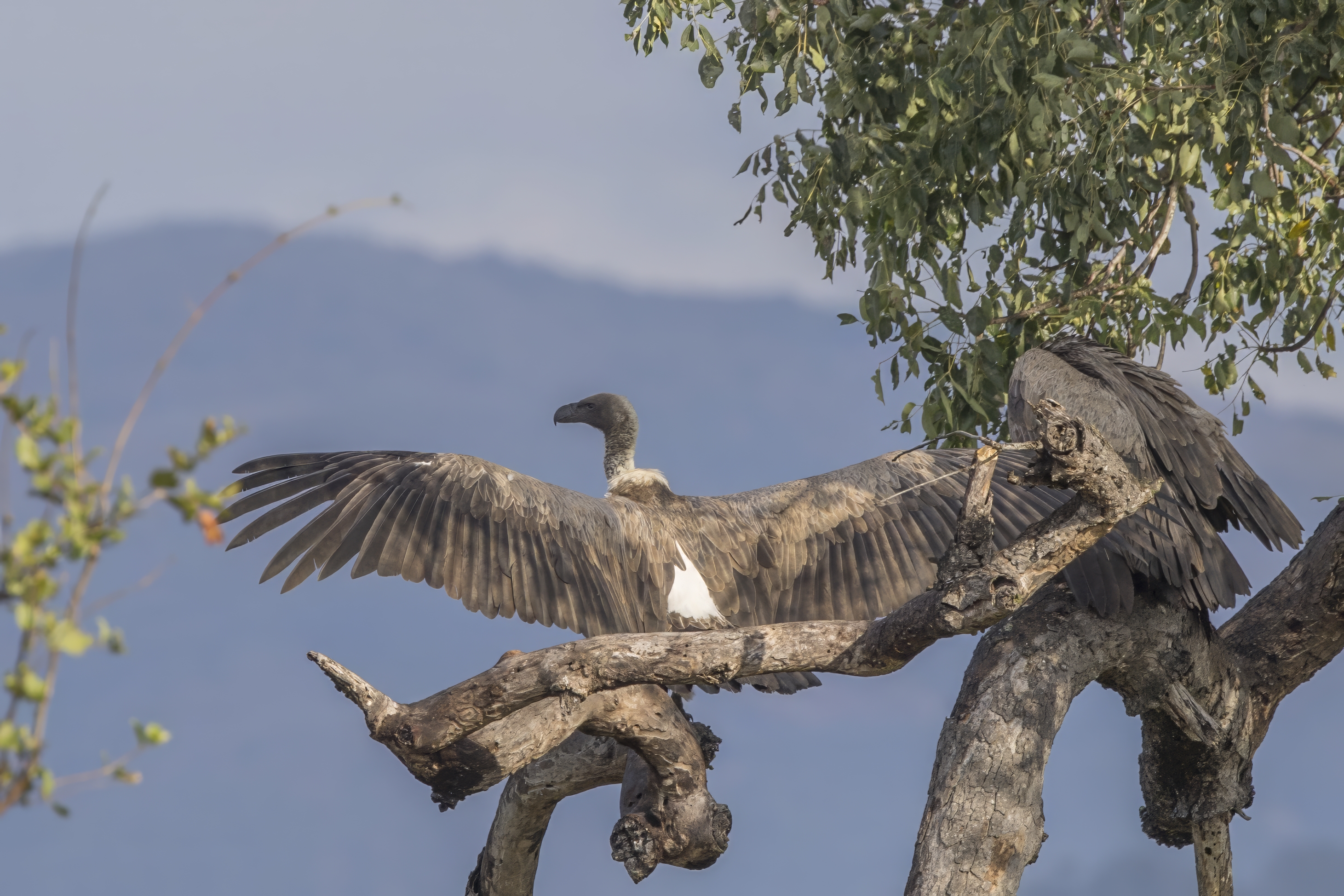
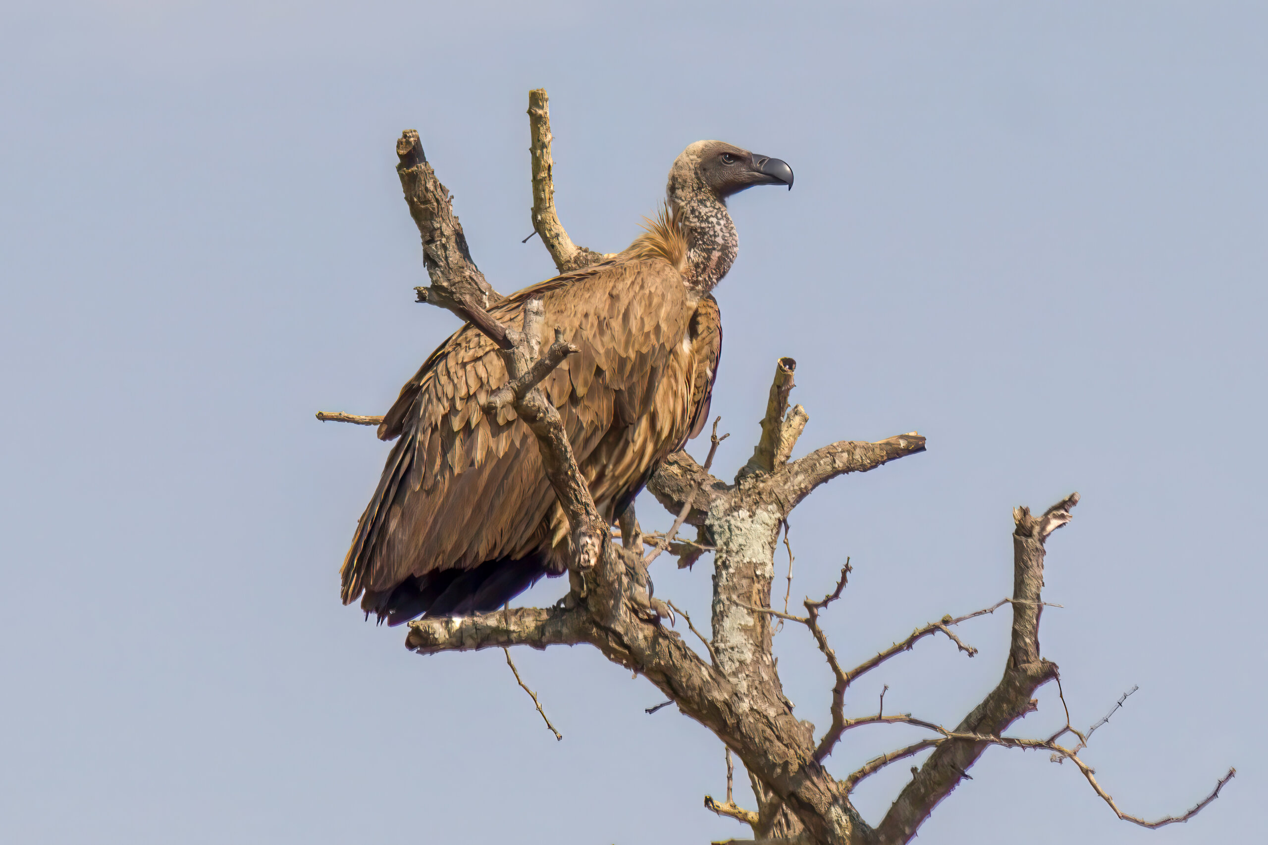
A bird’s organs sit within a compact body cavity, alongside an extensive respiratory system that includes air sacs. Seeing how these structures lie in relation to one another can be difficult with conventional views alone. In a preliminary study, researchers used contrast-enhanced computed tomography (CT) to examine the coelom—the body cavity containing the internal organs—of an adult male Gyps africanus, the White-backed Vulture.
The bird, a 22-year-old male living at Oasis Wildlife Fuerteventura in Spain, had been examined for an unrelated clinical condition. The researchers analysed those existing CT images. An iodine-based contrast agent given into a vein made blood vessels more visible, and views reconstructed from the scans revealed how organs, major vessels and air-filled structures lie alongside one another.
Different views revealed different structures
Soft-tissue settings made the heart, liver, spleen and kidneys particularly clear, while lung settings showed the lungs, air sacs and air-filled spaces within the skeleton more effectively. The contrast agent also revealed the aorta, which carries blood away from the heart, and major veins returning blood toward it. Among the visible vessels were the hepatic portal veins, which bring blood from digestive organs to the liver, and hepatic veins that carry blood away from the liver. Seeing these routes in three dimensions helps relate the circulatory system to the positions of the organs it supplies and drains.
The images did not resolve every structure. Individual sections of the intestine could not be distinguished reliably, and the boundaries of the air sacs could not consistently be followed. The researchers could not confidently identify individual parabronchi, the small passages within the lungs. The three-dimensional renderings also did not show the lungs, air sacs or gastrointestinal tract, which appeared with low attenuation in the scans.
The study offers a preliminary description of how contrast-enhanced CT can be used to examine the internal anatomy of a White-backed Vulture. The observed arrangement was broadly consistent with established avian anatomy, and the imaging approach may help inform future comparative and clinical studies. But the findings describe just one adult male, so they cannot establish the normal range of anatomical variation or provide reference values for the species. The CT identifications were also not checked against dissection or anatomical sections. The study is therefore a starting point for using these imaging methods, not a complete anatomical map of the species.
Anatomy & morphology
Related news
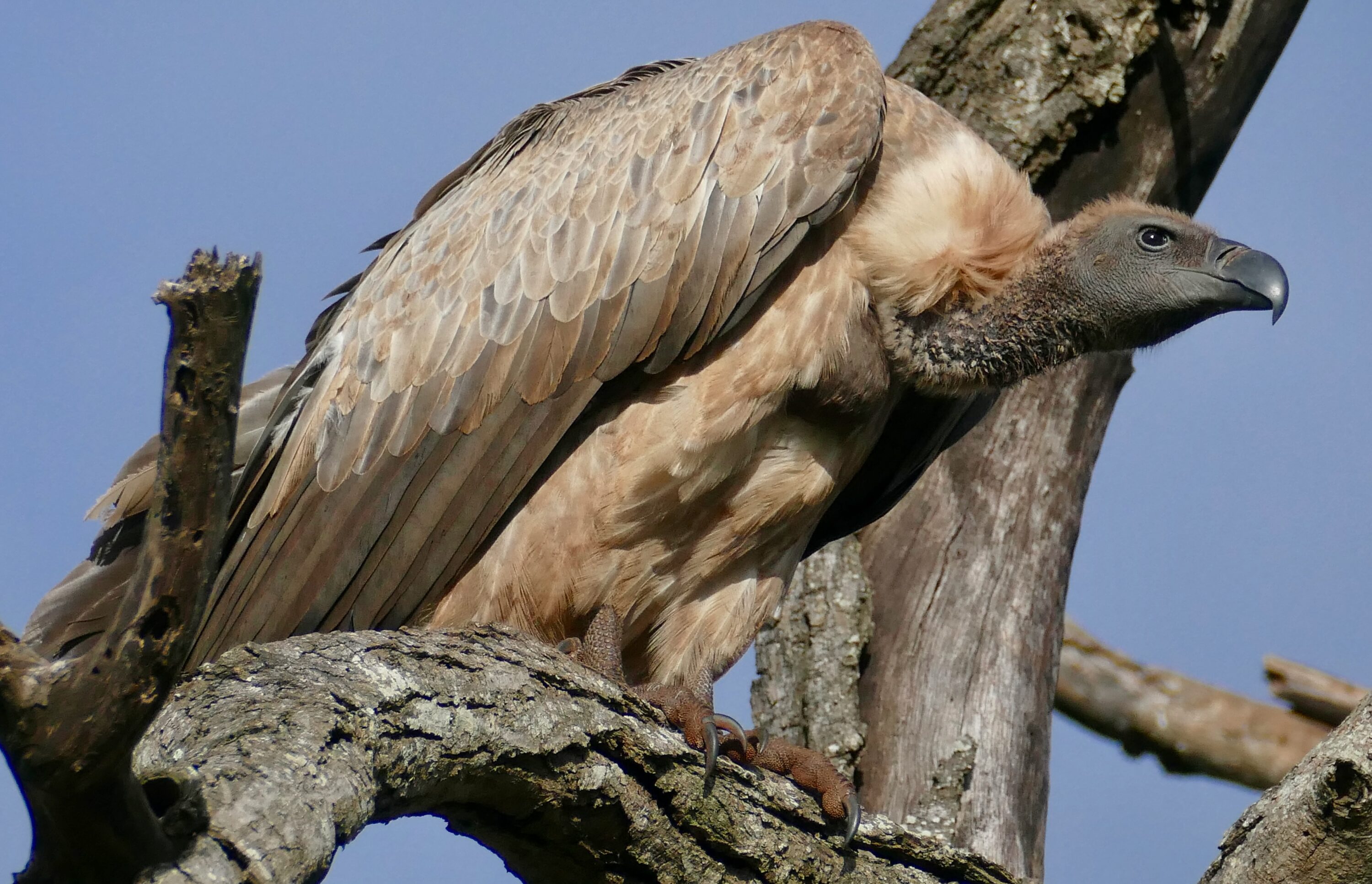
Two new studies sharpen the conservation map for white-backed vultures in Ethiopia
New surveys and habitat modelling from southwest Ethiopia identify important breeding, roosting and foraging areas for the critically…
Read story →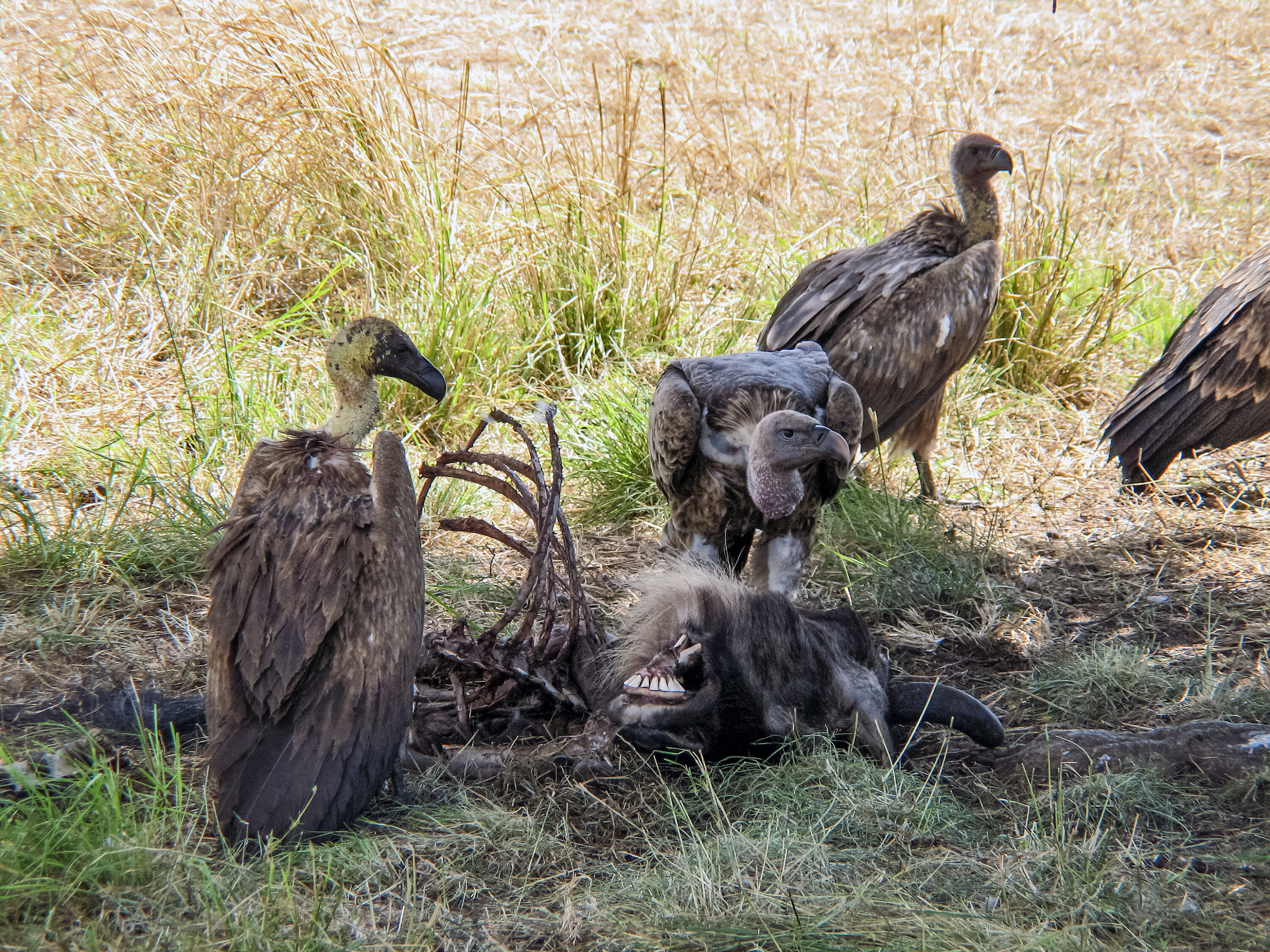
White-backed Vulture Transect Index Rose From 4.4 to 11.9 Birds per 100 km in Nazinga
Transect surveys in Burkina Faso’s Nazinga Game Ranch found 29 white-backed vultures in the latest survey and a…
Read story →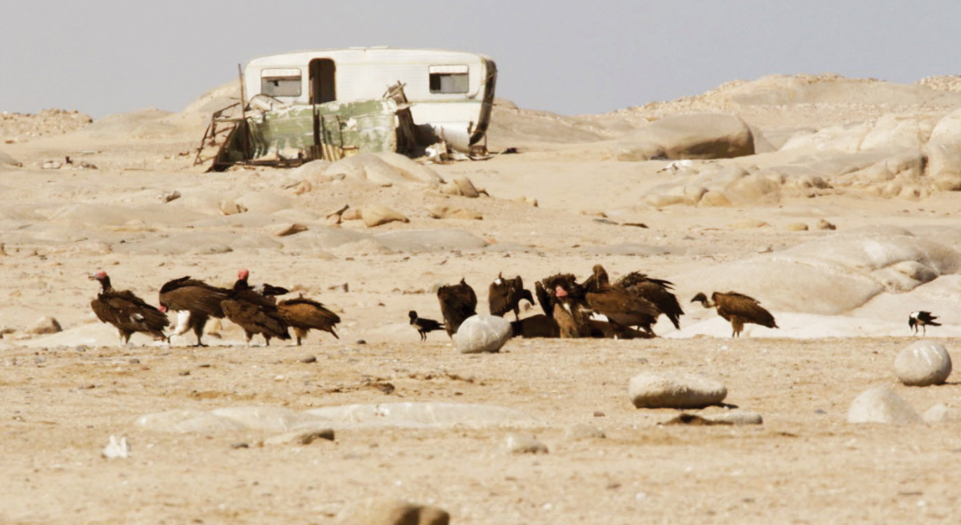
White-backed Vultures Documented Feeding on Marine Carrion for the First Time
Four immature white-backed vultures were observed feeding on a Cape fur seal killed by lions at Möwe Bay…
Read story →SPECIES IN THIS STORY
Species in this story
Independent research and conservation news archive
Did you find this information useful?
Wildlife Vagabond is independently built and maintained. Voluntary support helps cover source verification, hosting and continued work on research and conservation news.
Support Wildlife VagabondThe news archive will remain freely available.