
First Retinal Histology of Pied Crow Measured Total Thickness at About 303 µm
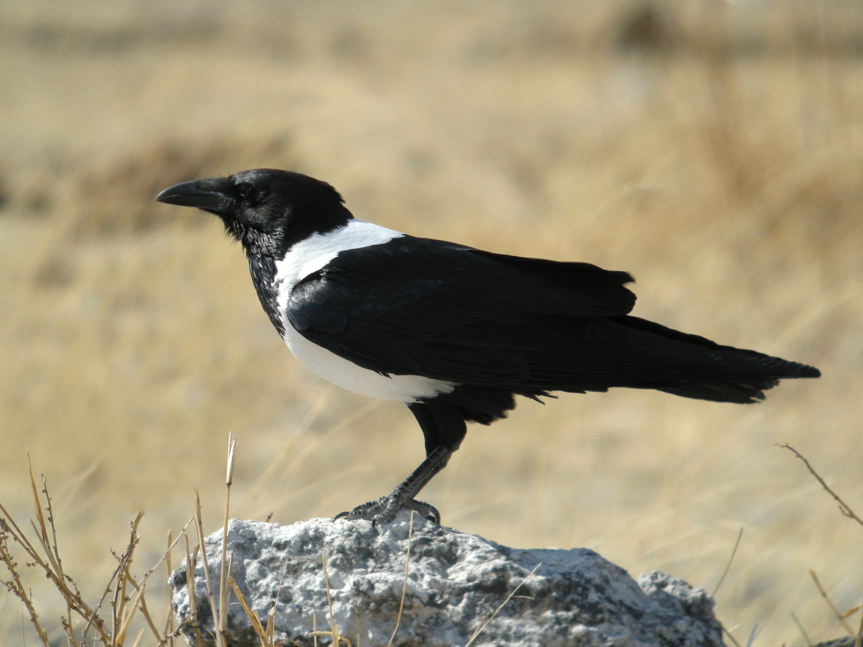
The first published histological description of the retina in the pied crow (Corvus albus) has documented the microscopic organisation of the species’ visual tissue. The study used both eyes from a single adult male obtained through an ethically approved wildlife surveillance programme in southwestern Nigeria, so the measurements are descriptive reference values rather than estimates for the species as a whole.
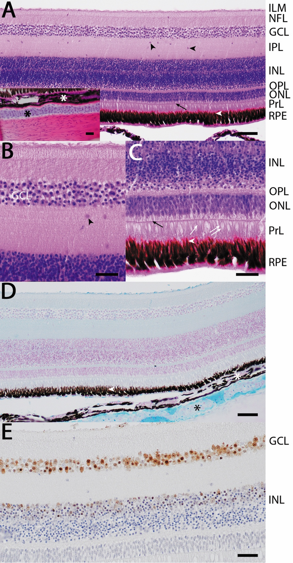
Researchers prepared thin tissue sections and used three complementary approaches. Hematoxylin and eosin staining showed the general retinal architecture, Alcian Blue highlighted the hyaline cartilage supporting the eye, and NeuN immunolabelling marked neuronal nuclei. NeuN was used to show where neurons were concentrated, not to identify specific neuronal subtypes.
The retina had the layered structure of a diurnal bird
The retina was organised into clearly defined layers typical of a visually guided diurnal bird. The outer nuclear layer contained three to four rows of nuclei and photoreceptors with morphologies consistent with both rods and cones. Beneath the retina, the choroid was densely pigmented and rich in blood vessels, while the sclera contained a prominent plate of hyaline cartilage that helps support the eye.
Total retinal thickness was about 303 µm
Total retinal thickness measured approximately 303 µm. The inner nuclear layer was about 80 µm thick, the inner plexiform layer about 58 µm, the outer nuclear layer about 25 µm and the ganglion cell layer about 27 µm. These values provide anatomical scale for this specimen rather than a population reference range.
NeuN labelling was strongest in the ganglion cell layer, whose neurons form a major output stage of the retina, and mild to moderate in the inner part of the inner nuclear layer. Within the sampled fields, the researchers counted about 25 NeuN-positive nuclei per 2,000 µm² in the ganglion cell layer. Around 20 measured NeuN-positive neuronal cell bodies ranged from 2.4 to 5.8 µm in diameter.
One adult male cannot describe variation within the species
The authors present the work as the first retinal histology dataset for Corvus albus and a baseline for future comparative studies of African corvid vision. Because only one opportunistically obtained adult male was available, the study cannot describe variation among individuals, sexes, ages or populations. Only one neuronal marker was used, different retinal regions were not compared, and possible foveal specialisations were not investigated.
Anatomy & morphology
Related news
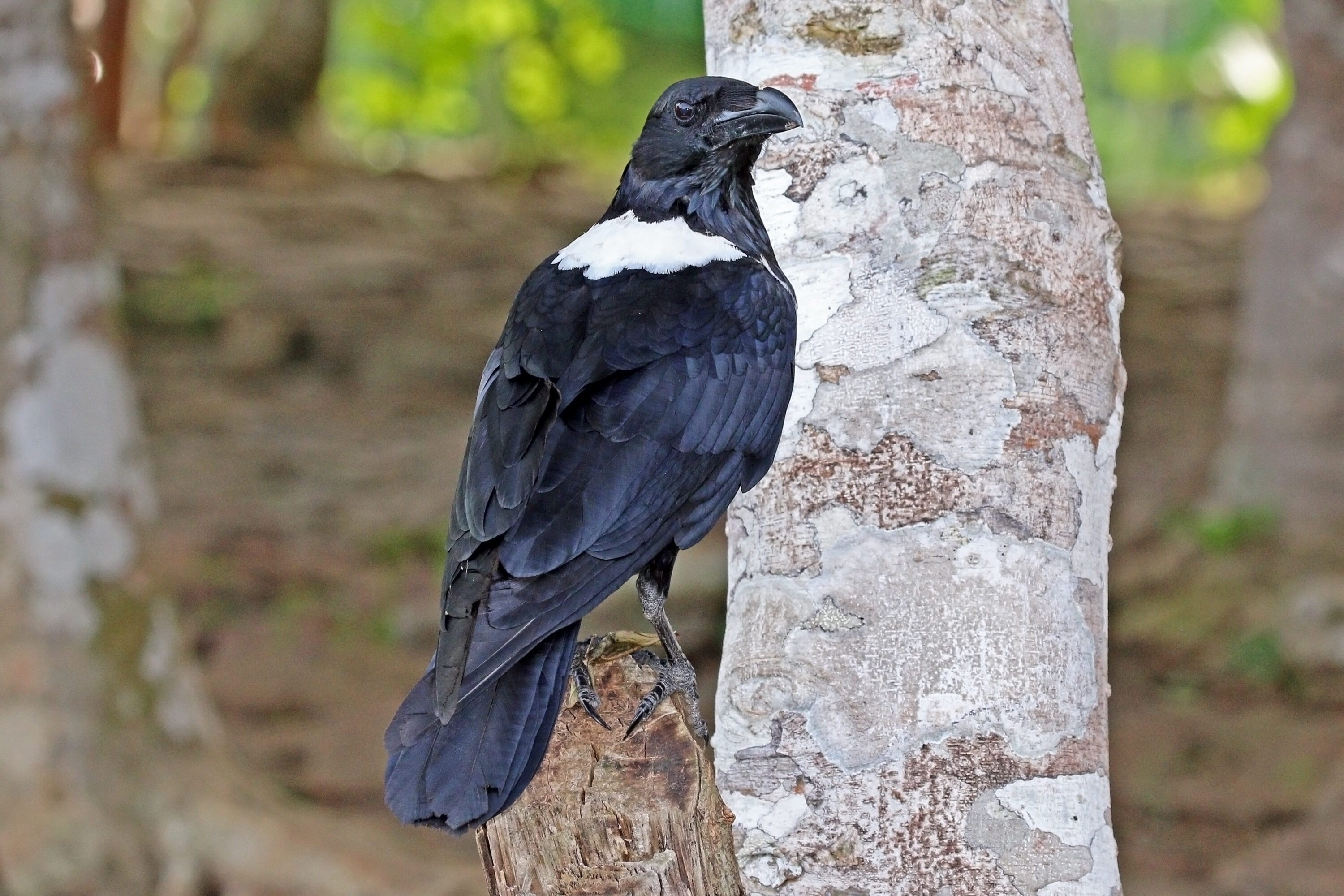
Pied Crow and Common Raven Began a Mixed Breeding Attempt at Jbel Moussa
A pied crow repeatedly seen with a common raven at Jbel Moussa in northern Morocco began a mixed…
Read story →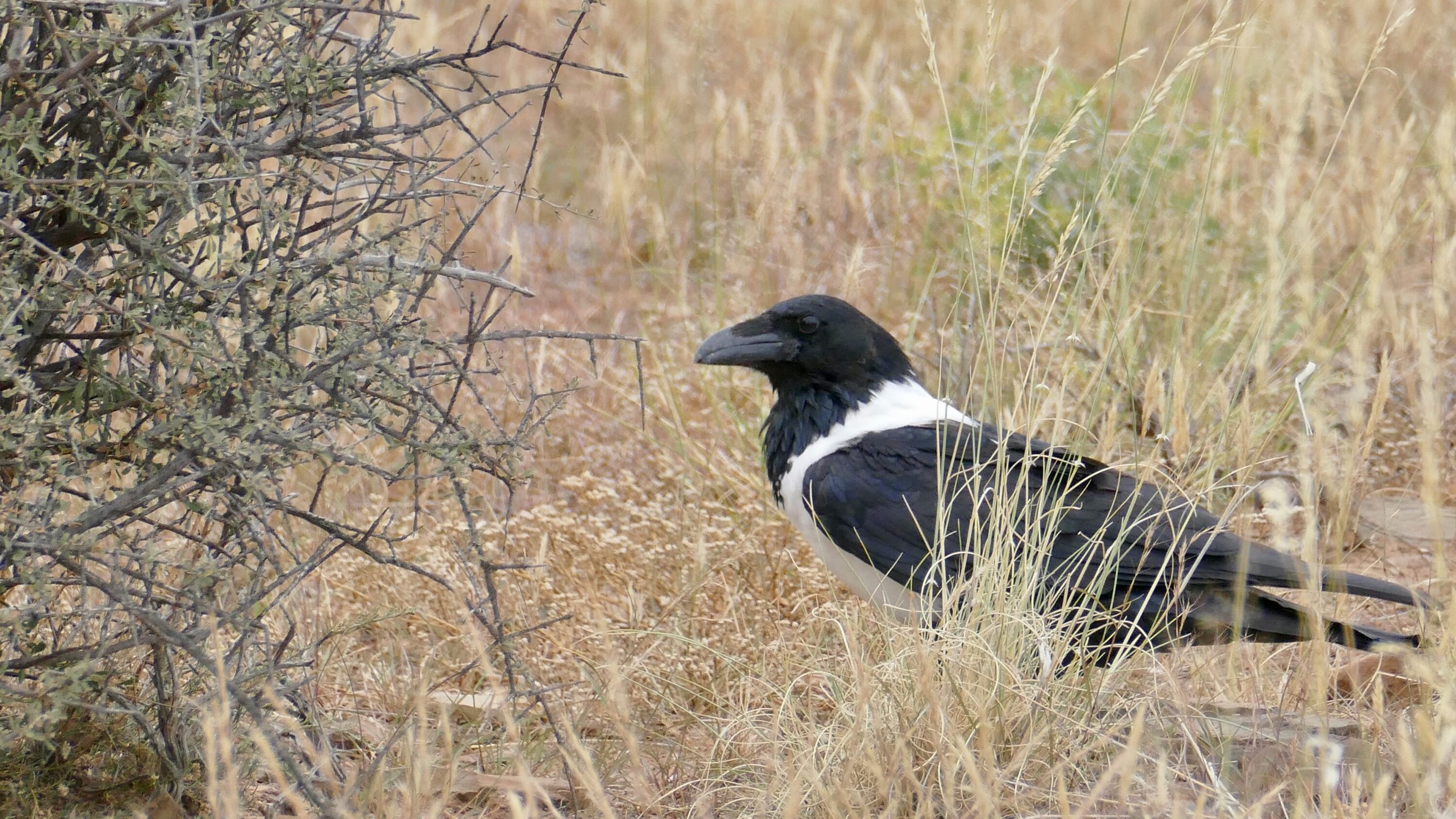
Britain’s Roaming Pied Crow Was Not Accepted as a Wild Vagrant
BOURC ruled in March 2026 that the pied crow roaming Britain in 2018–2019 should not be treated as…
Read story →
Pied Crows and Yellow-billed Kites Dominated Urbanized Ghanaian Shorelines in Both Seasons
Along a 15-km coast in Ghana’s Central Region, natural shorelines showed strong seasonal turnover in bird functional traits,…
Read story →SPECIES IN THIS STORY
Species in this story
Independent research and conservation news archive
Did you find this information useful?
Wildlife Vagabond is independently built and maintained. Voluntary support helps cover source verification, hosting and continued work on research and conservation news.
Support Wildlife VagabondThe news archive will remain freely available.