
Captive-raised Wild Boar Had a Denser Trabecular Network in the Talus
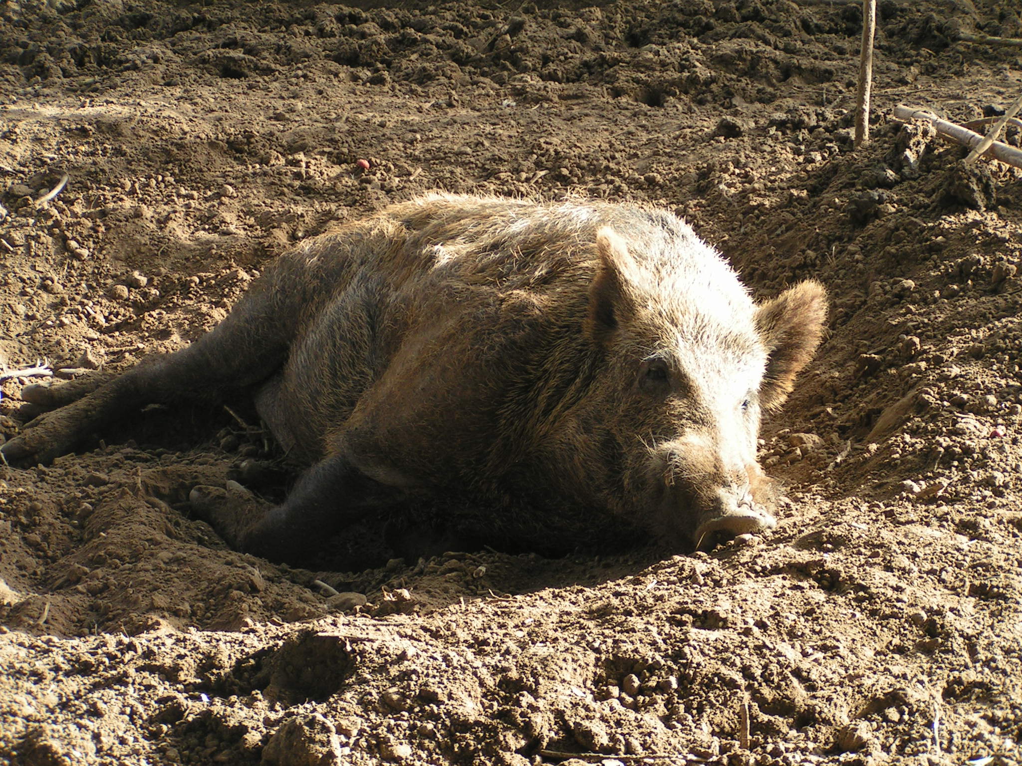
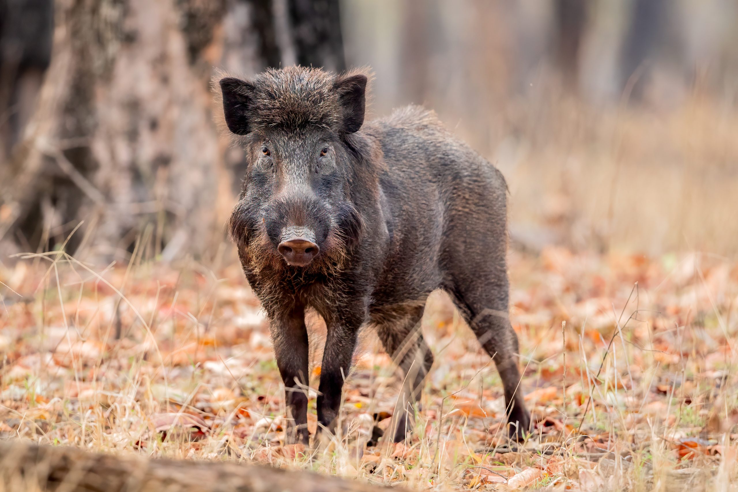
Wild boar (Sus scrofa) raised in captivity developed a different internal structure in the talus, the weight-bearing ankle bone, than animals that grew up in the wild. The 2026 French study compared 20 free-ranging boar with 21 wild-born animals raised from six months of age under reduced-mobility conditions: 10 in a large enclosure and 11 in a stable.
The researchers used CT-based three-dimensional maps and virtual sections to examine both the compact outer bone and the trabecular bone inside it. Trabecular bone is the lattice of small bony struts within the bone; a tighter network means that more of the internal space is occupied by closely packed load-bearing bone tissue.
Captive-raised boar had a denser internal trabecular network
The overall distribution of compact and trabecular bone remained similar across the groups, and the absolute thickness of compact bone changed little with mobility context. The captive-raised animals nevertheless had a denser, tighter trabecular network and a lower relative maximum thickness of compact bone. These quantitative differences remained after the researchers considered age, sex and body mass.
The authors therefore interpret the pattern as phenotypic plasticity: the same species can alter bone microanatomy during growth in response to the conditions it experiences, without a genetic change being required.
The result was the opposite of the original prediction
The researchers had expected reduced movement in captivity to lower mechanical loading and produce less trabecular and compact bone. Instead, the captive animals had denser trabecular bone. One possible explanation is that repetitive stereotyped movements or short bursts of activity in captivity created different mechanical stresses on the talus. The study did not demonstrate this mechanism, however, and did not find the clear directional trabecular pattern that such loading might be expected to produce.
All captive animals in the sample were the same age and were still growing when examined, so the study cannot show whether the same microanatomical pattern persists in older animals. The authors nevertheless suggest that plastic changes in talus microanatomy could become one line of evidence for detecting early stages of wild-ungulate domestication in archaeological remains.
Anatomy & morphology
Related news
Common Kestrels Had About Twice the Tracheal Mucous-Gland Density of White Storks
A comparative study of three adult males from each of three bird species found 11.32 tracheal mucous glands…
Read story →
Laughing Dove Eye Pecten Mapped From 12–13 Pleats to Cell Markers
Researchers mapped the pecten oculi, a vascular structure unique to bird eyes, in the laughing dove. Its 12–13…
Read story →
Progressive Greying Recorded in a Laughing Dove in Asia for the First Time
An adult laughing dove photographed in Karnataka, India, had localised white feathers on the head and shoulder region.…
Read story →SPECIES IN THIS STORY
Species in this story
Independent research and conservation news archive
Did you find this information useful?
Wildlife Vagabond is independently built and maintained. Voluntary support helps cover source verification, hosting and continued work on research and conservation news.
Support Wildlife VagabondThe news archive will remain freely available.