
Laughing Dove Eye Pecten Mapped From 12–13 Pleats to Cell Markers
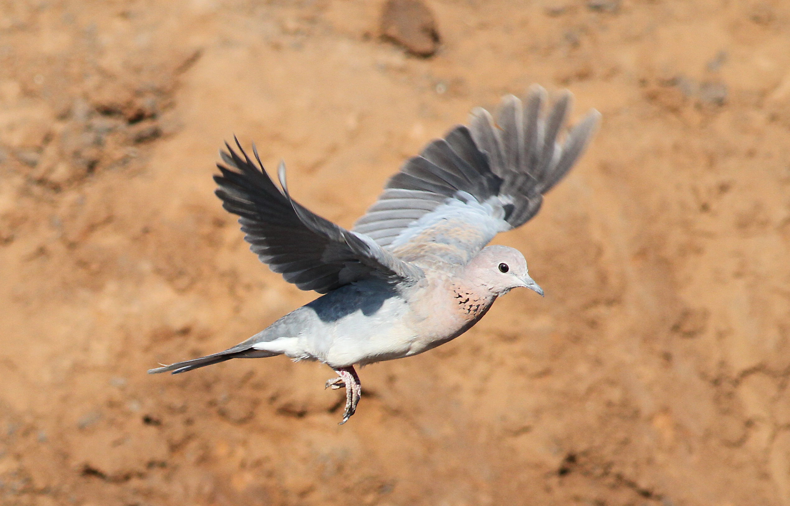
The pecten oculi is a specialised, highly vascular structure found only in bird eyes. A study published on 9 September 2026 mapped this structure in the laughing dove (Spilopelia senegalensis) from its overall folds down to cell types, structural proteins and elemental composition. The researchers combined light microscopy, scanning electron microscopy, energy-dispersive X-ray microanalysis and immunofluorescence with ten antibodies.
From 12–13 pleats to elemental differences
The pecten was the pleated type, with a basal part, 12–13 folded pleats and bridges connecting them near the apex. Its folded surfaces were lined by capillaries and the vitreopectineal limiting membrane. Between the pleats, the researchers found pigment cells and amoeboid hyalocytes, while light microscopy showed vascular channels surrounded by melanocytes and hyalocytes.
Energy-dispersive X-ray microanalysis, which identifies elements present in a tissue sample, showed that the pecten was not chemically uniform. Carbon, oxygen and nitrogen dominated overall, but sodium was detected at the apex, fluorine in the middle pleats and magnesium at the base.
Antibodies mapped different cells and supporting tissues
The immunofluorescence markers added a cellular map to the anatomy. CD34 and vimentin marked endothelial cells and parts of the limiting membrane, while PDGFRα occurred in the limiting membrane and in cells associated with capillaries. Melan-A and SOX10 identified melanocytes and other pigment-associated cells around the capillaries, while CD45 marked macrophage-like cells in the interstitial tissue.
Collagen I and III and laminin mapped supporting extracellular and basement-membrane structures. Tom20 marked mitochondria within the blood capillaries, while α-SMA was present in the pleats, base, capillaries and interstitial tissue. Together, the results show that the laughing dove pecten is a highly organised structure in which blood vessels, pigment cells, immune-like cells and supporting tissues are distributed in distinct patterns. The authors present the study as an anatomical and immunophenotypic reference for future comparisons of the pecten across birds.
Anatomy & morphology
Related news
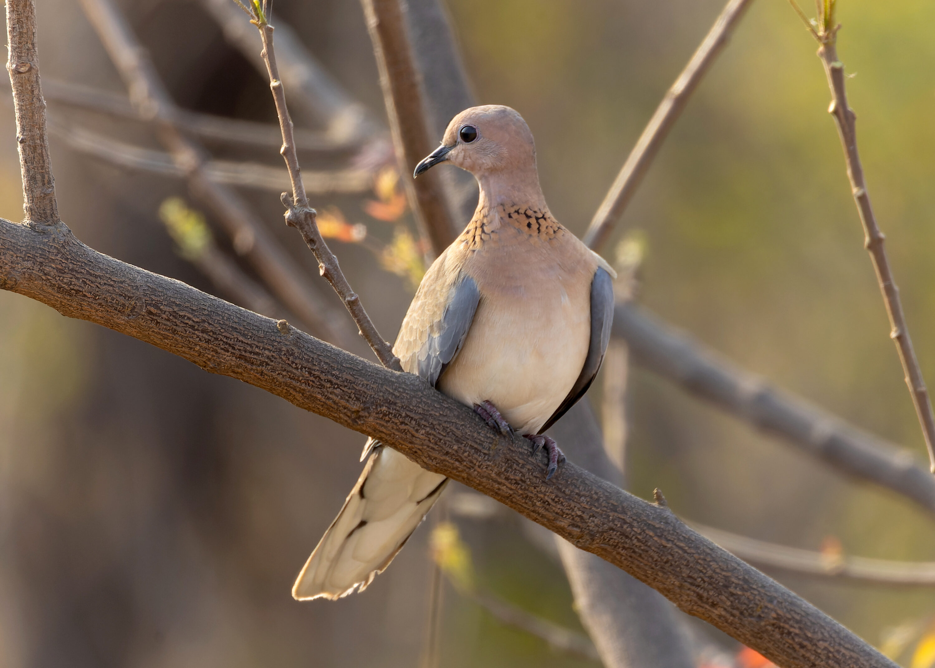
Progressive Greying Recorded in a Laughing Dove in Asia for the First Time
An adult laughing dove photographed in Karnataka, India, had localised white feathers on the head and shoulder region.…
Read story →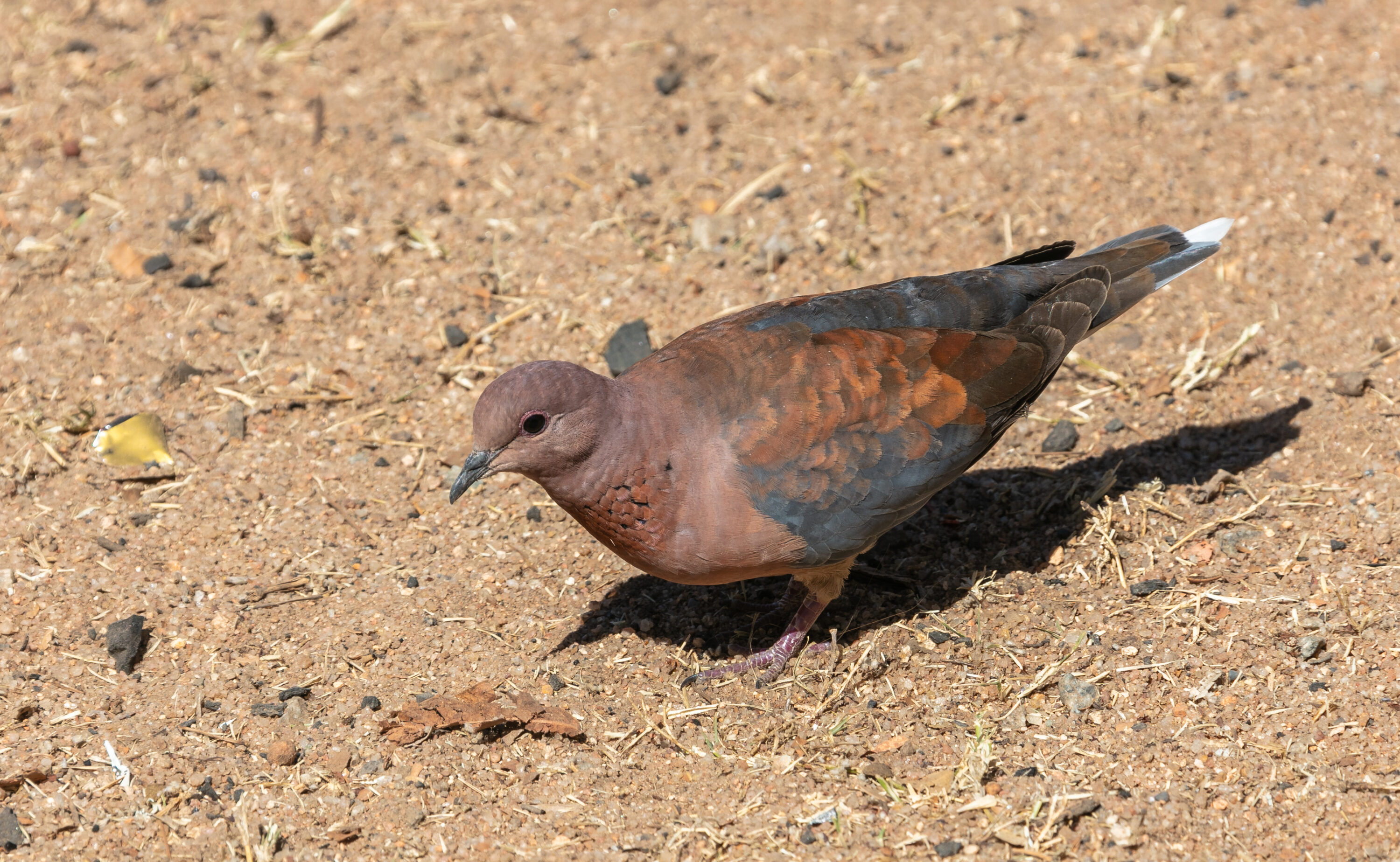
Laughing Dove Preen Secretions Showed Strong Antimicrobial Activity
A 2026 comparative study found that laughing doves had the thinnest epidermis of three bird species and that…
Read story →
South African Nest Records Revealed Species-specific Shifts in Breeding Timing
A 2026 thesis using thousands of South African nest records found no single national shift in breeding timing:…
Read story →SPECIES IN THIS STORY
Species in this story
Independent research and conservation news archive
Did you find this information useful?
Wildlife Vagabond is independently built and maintained. Voluntary support helps cover source verification, hosting and continued work on research and conservation news.
Support Wildlife VagabondThe news archive will remain freely available.