
Congenital heart defect discovered in lion cub during severe illness
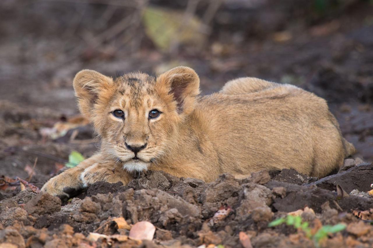
A 137-day-old female lion (Panthera leo) in Jamnagar, India, was brought for treatment with severe weakness, poor coordination, difficulty breathing and dehydration. Blood tests showed dangerously low blood sugar and signs of damage affecting several organs. An elevated level of cardiac troponin I, a marker of heart-muscle injury, prompted an echocardiogram after her condition had been stabilized.
The scan revealed a 2.2-centimetre opening in the lower part of the wall between the heart’s two atria, close to the valves separating the upper and lower chambers. The researchers classified it as an incomplete atrioventricular septal defect (AVSD). AVSDs are congenital defects involving the central junction where the heart’s chambers and atrioventricular valves meet. In an incomplete form, the abnormal passage is between the atria rather than between both the atria and ventricles.
Normally, blood flows from the right atrium to the right ventricle and then to the lungs; blood returning from the lungs enters the left atrium. Color Doppler imaging showed that most blood crossing the opening flowed from left to right. This sends extra blood back through the right side of the heart, increasing its workload. The scan also showed mitral valve regurgitation—some blood leaking backward through the valve on the left side of the heart—although its severity was not measured.
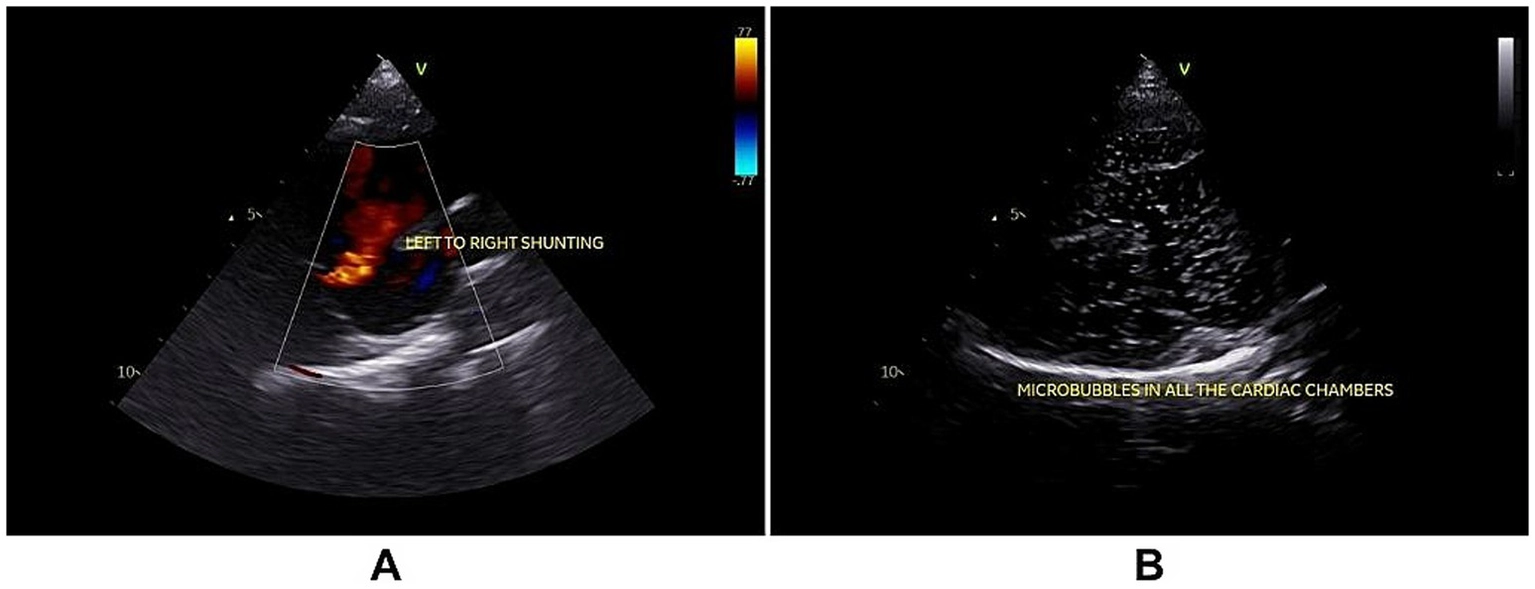
The saline-contrast test showed microbubbles passing to the left side of the heart, indicating an additional right-to-left component to the shunt. The researchers performed the heart imaging under anesthesia while the cub was ill; they note that these conditions can influence blood flow and the apparent direction of a shunt. The cub’s blood sugar returned to normal with treatment, but kidney problems and worsening bilirubin levels developed, and she died on the fourth day.
Examination after death confirmed the defect, measuring about 2 by 2.5 centimetres, and found excess volume on the right side of the heart and fibrosis—scar-like tissue—in the right ventricle. Together, the right heart’s volume overload and the fibrosis show that the shunt was hemodynamically significant and had been present long enough to affect the heart. The authors considered the defect incidental to the acute illness, whose cause remained unknown. They could not determine whether it contributed to the cub’s death.
The authors describe this as the first reported AVSD in a lion and, to their knowledge, in any species of Panthera. A single case cannot show how common the defect is, but it documents its anatomy and its effects on one lion’s heart.
Anatomy & morphology
Related news
A lion liver protein was a poor hepatitis B virus receptor—and a less efficient bile-acid transporter
Tests of NTCP proteins from cats, tigers and leopards supported hepatitis B virus binding, unlike lion NTCP, which…
Read story →
A graph-based tool helps researchers match wildlife photos to individual animals
A new method combines automated photo matching with targeted human review. Across seven wildlife image datasets, it improved…
Read story →
A Little-Known Microbial World Inside Lions’ Guts
Faecal samples from wild lions in Namibia gave researchers a much clearer picture of their gut bacteria. Microbial…
Read story →SPECIES IN THIS STORY
Species in this story
Independent research and conservation news archive
Did you find this information useful?
Wildlife Vagabond is independently built and maintained. Voluntary support helps cover source verification, hosting and continued work on research and conservation news.
Support Wildlife VagabondThe news archive will remain freely available.