
Study Mapped Green-winged Teal Lung Architecture From Bronchi to Air Capillaries
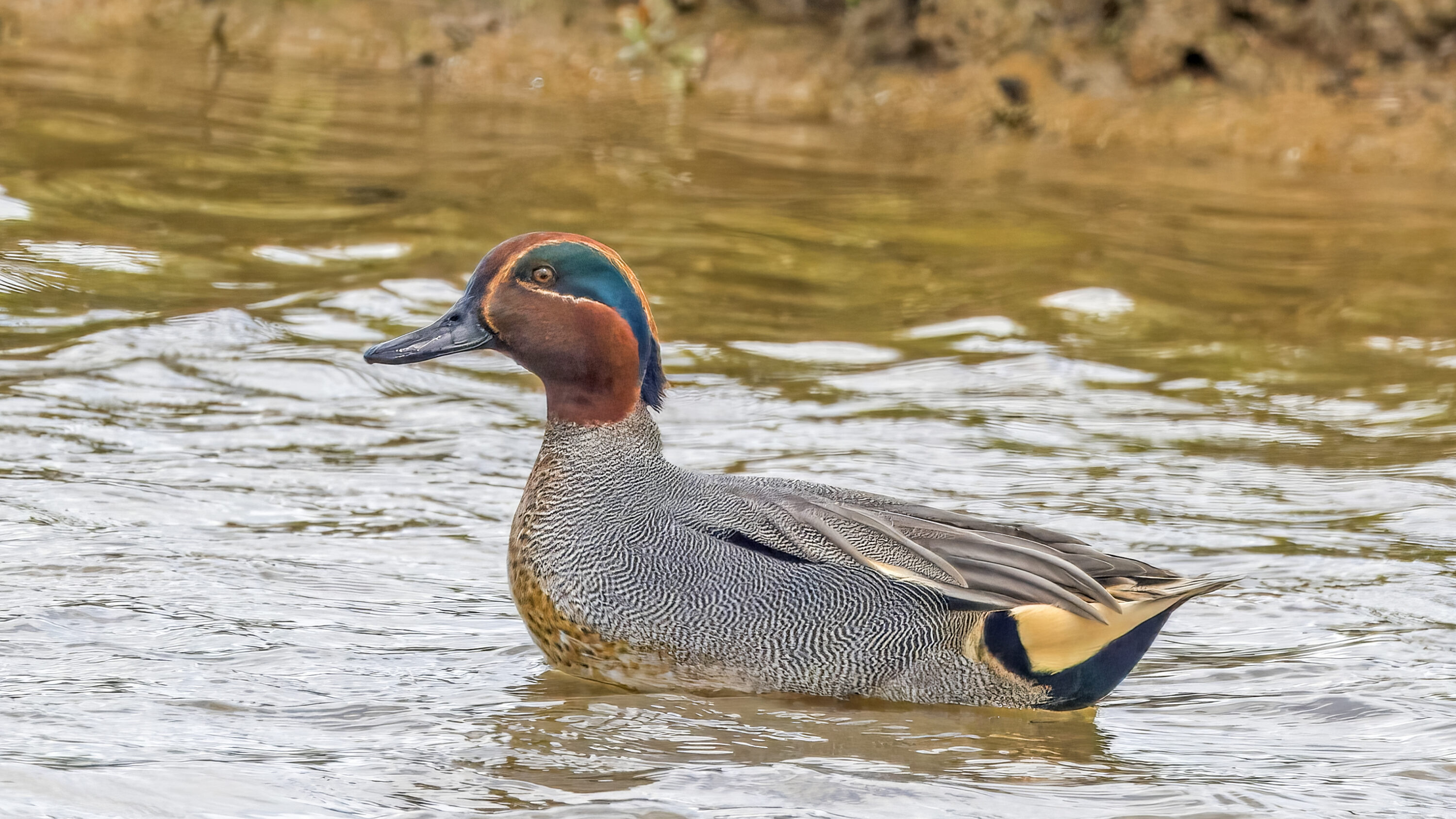
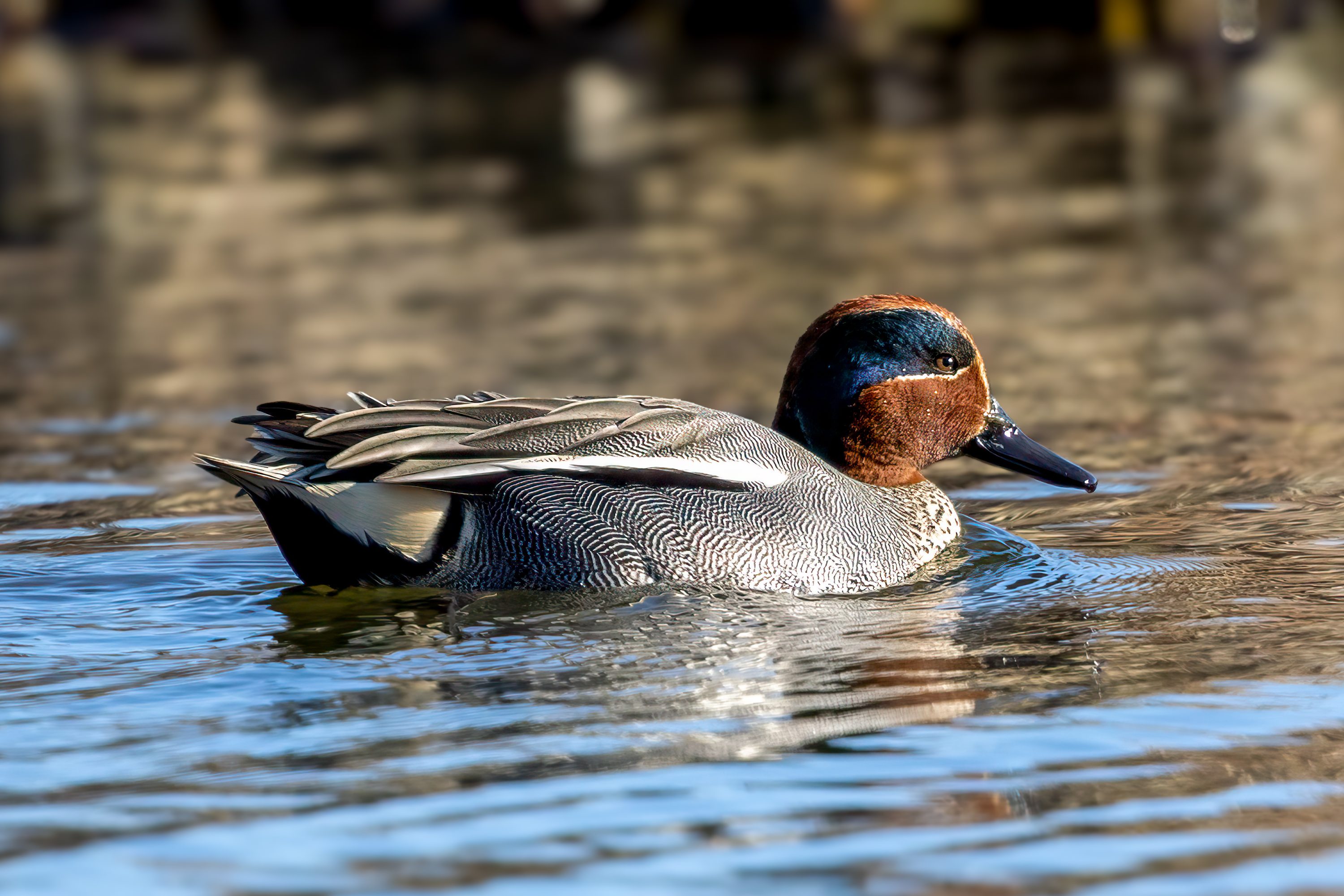
A 2026 study described the lungs of Green-winged Teal (Anas crecca) from whole-organ anatomy down to the microscopic passages involved in moving air through the avian lung. Researchers examined lungs from 20 healthy adults collected after slaughter in Kafr El-Sheikh Governorate, Egypt, using dissection, cross-sections, corrosion casts, histology and scanning electron microscopy.
The lungs were trapezoidal and non-lobated and fitted tightly against the ribs in the craniodorsal part of the body cavity — toward the back and headward side of the torso. This differs from the soft, lobed lungs familiar from mammals: bird lungs are relatively rigid, while air is routed through a branching bronchial system and the associated air sacs.
Secondary bronchi fed a network of parabronchi
Corrosion casts showed how the intrapulmonary primary bronchus branched into several groups of secondary bronchi. The teal consistently had seven laterodorsal and four lateroventral secondary bronchi, with additional medioventral and posterior groups. These names describe the direction of the branches: laterodorsal bronchi run toward the outer and dorsal side of the lung, lateroventral toward the outer and ventral side, and medioventral toward the midline and ventral side. The posterior branches were shortest and the medioventral group largest.
The biological point of this branching pattern is what comes next. Secondary bronchi are connected by parabronchi — narrow tubes through which air moves inside the lung. The paleopulmonic and neopulmonic regions are two structural parts of this parabronchial system. In the paleopulmonic region the parabronchi were elongated, whereas in the neopulmonic region they formed a more hexagonal, network-like arrangement.
Atria opened toward the fine air-capillary system
Scanning electron microscopy showed numerous atrial openings in the walls of the parabronchi. The atria are small chambers opening from the parabronchi toward still finer passages and the air-capillary system, where air is brought into close contact with blood capillaries for gas exchange. Histology also showed ciliated lining in the primary and secondary bronchi, smooth-muscle layers and connective-tissue septa supporting the parabronchial organisation.
The study therefore provides a species-specific anatomical map rather than a direct measurement of respiratory performance. The authors present it as baseline material for comparing lung structure among waterfowl and for future work linking anatomical differences to respiratory function.
Anatomy & morphology
Related news
Seven overlaps linked wild-bird H5 clusters with poultry outbreaks in the US
Across seven periods between 2022 and 2025, clusters of H5 detections in wild waterfowl overlapped in time and…
Read story →
Mallard Population Growth Was Lower in Years With Longer Growing Seasons at Higher Latitudes
Across 38 years of western boreal survey data, annual mallard population growth was lower in years with longer…
Read story →
Green-winged Teal With Plastic Nasal Saddles Had Lower Estimated Annual Survival in a Study of 11,874 Birds
A 20-year French marking dataset found lower estimated annual survival in Green-winged Teal fitted with plastic nasal saddles…
Read story →SPECIES IN THIS STORY
Species in this story
Independent research and conservation news archive
Did you find this information useful?
Wildlife Vagabond is independently built and maintained. Voluntary support helps cover source verification, hosting and continued work on research and conservation news.
Support Wildlife VagabondThe news archive will remain freely available.