
H5N1 Targets the Brain and Eyes of Black-headed Gulls
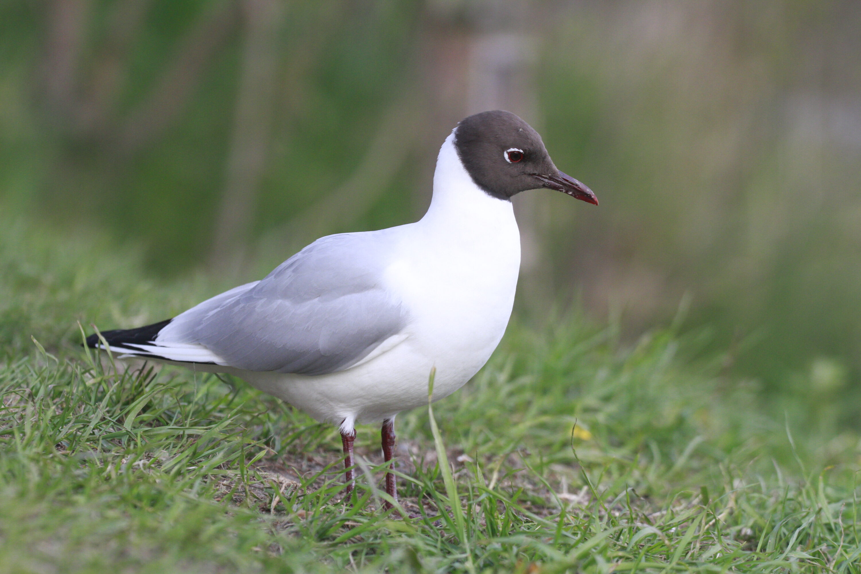
A study of naturally infected wild black-headed gulls (Chroicocephalus ridibundus) in France found that H5N1 high pathogenicity avian influenza showed a strong affinity for the nervous system and consistently involved structures inside the eye. H5N1 is a subtype of influenza A virus; the birds carried clade 2.3.4.4b, the H5 lineage behind major recent outbreaks and mortality in European wild birds. “High pathogenicity” is an avian-influenza classification based on the capacity to cause severe disease in poultry; disease severity can differ among wild bird species. The study was published in Scientific Reports on 8 June 2026.
Between December 2022 and February 2023, 49 black-headed gulls from northern France tested positive for H5N1. Thirty-seven were admitted alive for diagnostic investigation and 12 were found dead in the field. The birds were free-living wild gulls and were not part of an experimental infection study. Researchers selected 13 of the 49 cases for detailed pathological analysis based on the condition of the carcasses and tissues. They examined tissue microscopically for lesions, used RT-qPCR to detect viral RNA, and used immunohistochemistry — antibodies that stain influenza proteins in tissue sections — to show which organs and cell types contained viral antigen. RNA detection shows that viral genetic material is present; antigen inside damaged cells provides more direct anatomical evidence that the tissue itself was involved in the infection.
Brain lesions were found in all 13 gulls
Clinical information was available for 36 live birds received by the Wildlife Care Center of the National Veterinary College of Alfort. Twenty-five were emaciated and 18 showed neurological signs, including altered alertness, head tremors, abnormal head posture and other movement abnormalities. Seven had conjunctivitis with excessive tearing.
All 13 gulls selected for detailed pathology had encephalitis, and viral antigen was detected in the brain of all 13. The cerebrum was consistently involved, while the ventricular system and midbrain were also frequently positive. This is marked neurotropism: the virus preferentially involved nervous tissue, with antigen concentrated mainly in neurons and supporting glial cells. By contrast, the researchers found no viral antigen in endothelial cells lining blood vessels, so the pattern was not one of endotheliotropism.
Viral antigen was also found in other organs. After the brain, the eye and pancreas were the most frequently positive tissues: antigen was detected in the eye in 10 of 13 gulls and in the pancreas in 8 of 13.
The eye was a direct target of infection
Ocular lesions were common among the 13 gulls. Ten had endophthalmitis — inflammation involving tissues inside the eye — and recurring changes included anterior uveitis, inflammation of the front part of the vascular middle layer of the eye, and retinal degeneration. Viral antigen was detected in several ocular structures, especially the trabecular meshwork, which helps drain fluid from the front of the eye, and the retina. The optic nerve remained negative.
Earlier field observations had described post-H5N1 eye abnormalities in some marine birds, but comparable ocular changes had not previously been documented in gulls in this way. The combination of tissue damage, immunohistochemical detection and viral RNA led the authors to identify the eye as an underestimated target organ in H5N1 infection.
The study describes selected confirmed cases
The 13 birds were selected from confirmed cases because their tissues were sufficiently well preserved for detailed examination. The study therefore describes the pathology and tissue distribution of H5N1 in infected gulls rather than estimating how common infection or severe disease was in the wider population.
The authors also detected viral antigen in feather epithelium and, in some birds, the uropygial or preen gland at the base of the tail. This suggests that skin-, feather- and preen-gland tissues could contribute to viral shedding, although the study did not directly measure how much infectious virus these tissues released. The authors caution that the work was observational and based on a limited number of naturally occurring cases, so the pattern may not apply to every H5N1 strain or epidemiological setting.
Overall, the selected infected gulls showed neurotropism and oculotropism — preferential involvement of nervous and eye tissues — together with selective infection of some epithelial tissues, but no preference for the endothelial cells lining blood vessels. The authors therefore recommend including the brain, eyes and skin- and feather-related tissues in HPAI surveillance and diagnostic work in marine birds.
Disease, parasites & microbiology
Related news
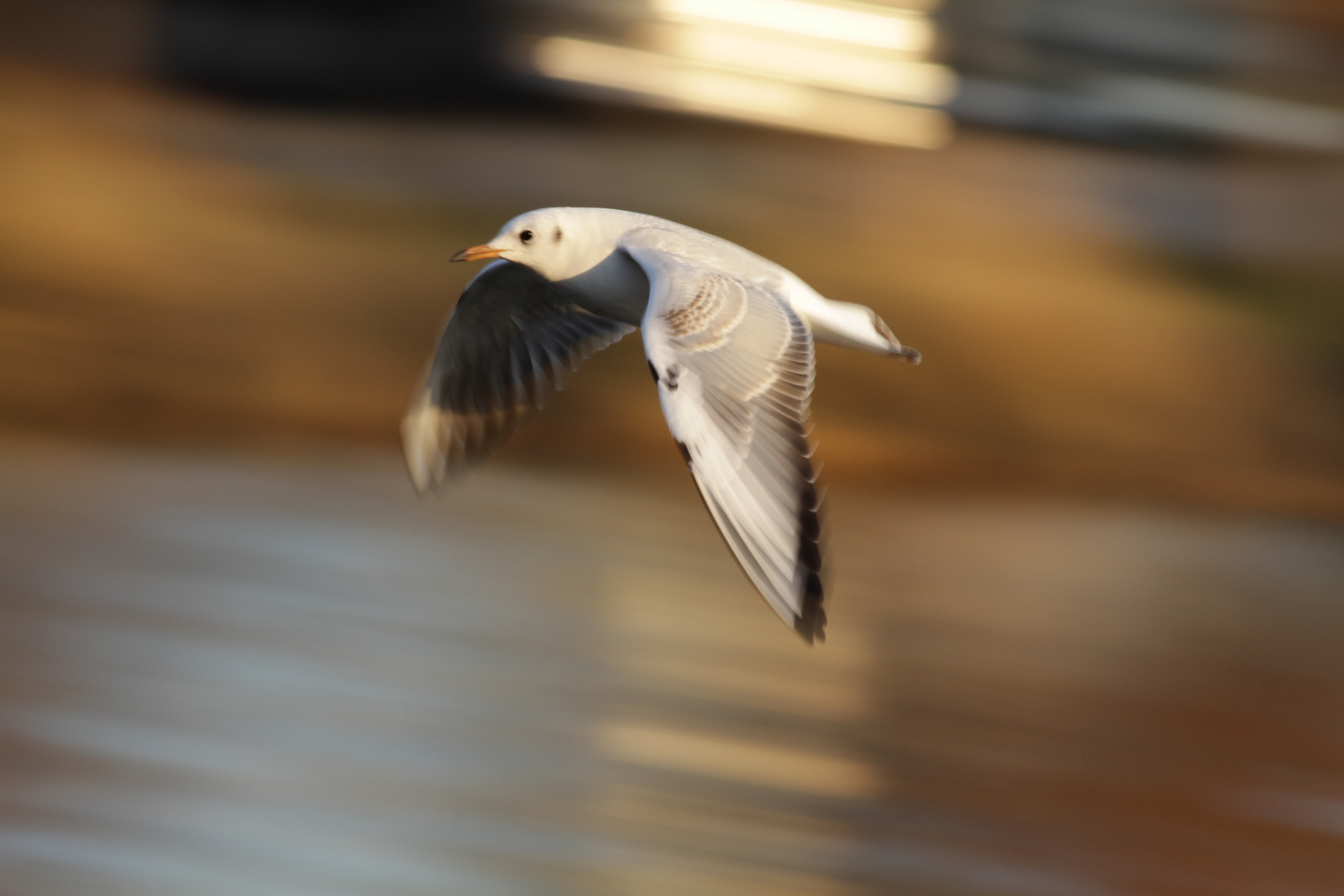
GPS-Based Models Highlighted the Lower Sumida as a Key Black-headed Gull Corridor in Tokyo
GPS tracking showed that black-headed gulls followed different river features at different movement scales in Tokyo, while GPS-based…
Read story →
Black-headed Gull Sample Yielded the Longest Known Calicivirus Genome
A Hungarian black-headed gull faecal sample yielded an 8,845-nucleotide avian calicivirus genome with unusual RNA structures and enough…
Read story →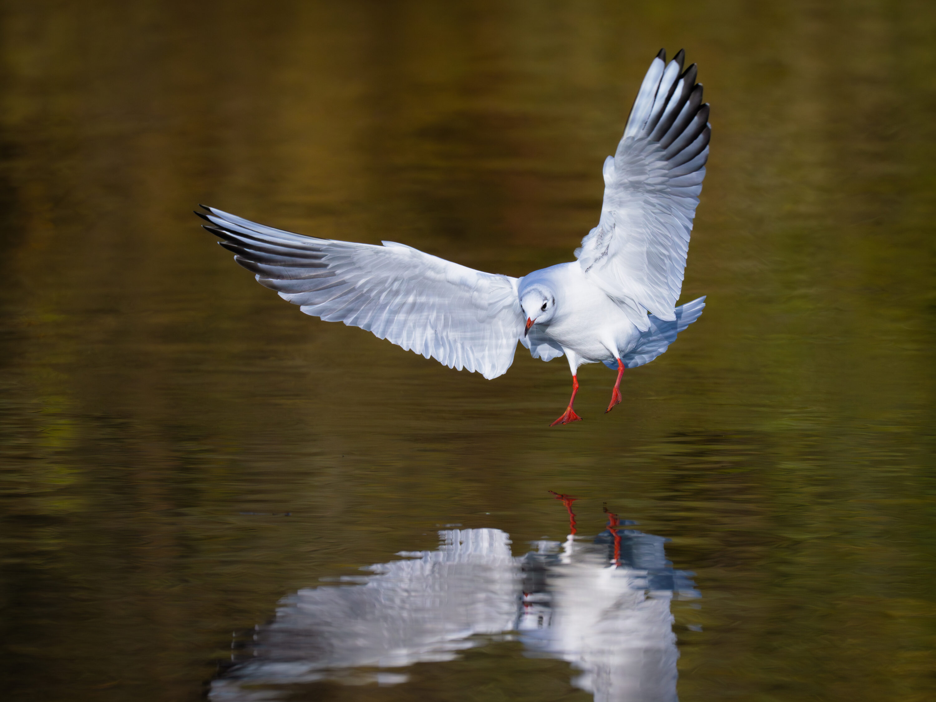
New Papillomavirus Genome Identified in Black-headed Gull Faeces in China
Researchers identified the 7,794-base-pair CrPV1 genome in faecal samples from black-headed gulls in Sichuan and found it genetically…
Read story →SPECIES IN THIS STORY
Species in this story
Independent research and conservation news archive
Did you find this information useful?
Wildlife Vagabond is independently built and maintained. Voluntary support helps cover source verification, hosting and continued work on research and conservation news.
Support Wildlife VagabondThe news archive will remain freely available.