
Microscopy Study Mapped the Upper Digestive Tract of Adult Spur-Winged Geese
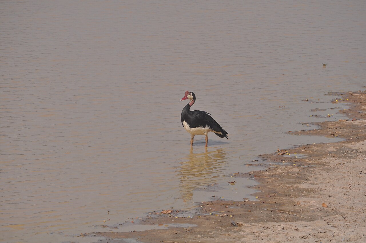
A microscopic study of the upper digestive tract in spur-winged geese (Plectropterus gambensis) has provided a detailed description of tissues from the beak to the gizzard and the beginning of the small intestine. The study was published on 15 March 2026 in Folia Veterinaria.
Researchers examined six apparently healthy adult birds, three males and three females, purchased from local sellers in Borno State in northeastern Nigeria. Tissue samples from the beak, tongue, oesophagus, proventriculus and gizzard were prepared for light microscopy, with additional staining used to examine mucus-producing tissues.
Beak and tongue combined keratinisation, sensory structures and mucus glands
The beak was covered by keratinised stratified squamous epithelium. Beneath it, the researchers found dense connective tissue, blood vessels and encapsulated sensory nerve endings known as Herbst corpuscles. The authors describe these as pressure-sensitive mechanoreceptors that can help a waterbird detect pressure and vibration while selecting and assessing food. The underside of the tip of the tongue was also strongly keratinised, while the tongue contained extensive compound tubular mucus glands.
The oesophagus was lined by non-keratinised stratified squamous epithelium. Beneath this lining were mucus glands and small lymphatic nodules. The authors note that mucus from these glands lubricates the passage of food and protects the epithelial surface against friction. At the transition from the oesophagus to the proventriculus, the mucosal glands formed crypt-like folds.
Glandular stomach and gizzard showed distinct digestive specialisations
Much of the wall of the proventriculus consisted of glands lined by oxynticopeptic cells, the avian cells involved in secreting both acid and digestive enzymes. At the isthmus between the proventriculus and gizzard, the proventricular folds transitioned into organised gizzard glands.
The gizzard itself had a hard cuticle covering its inner surface and a well-developed muscular wall. The authors relate this muscular construction to the gizzard’s role in grinding and macerating tough food. At the transition from the gizzard to the duodenum, the researchers recorded mucosal folds of varying height, lymphatic nodules and the first intestinal villi.
The authors present the study as a detailed anatomical reference for the species. They note that such information can support comparisons among waterfowl and help relate the structure of the digestive tract to feeding adaptations in spur-winged geese.
Anatomy & morphology
Related news
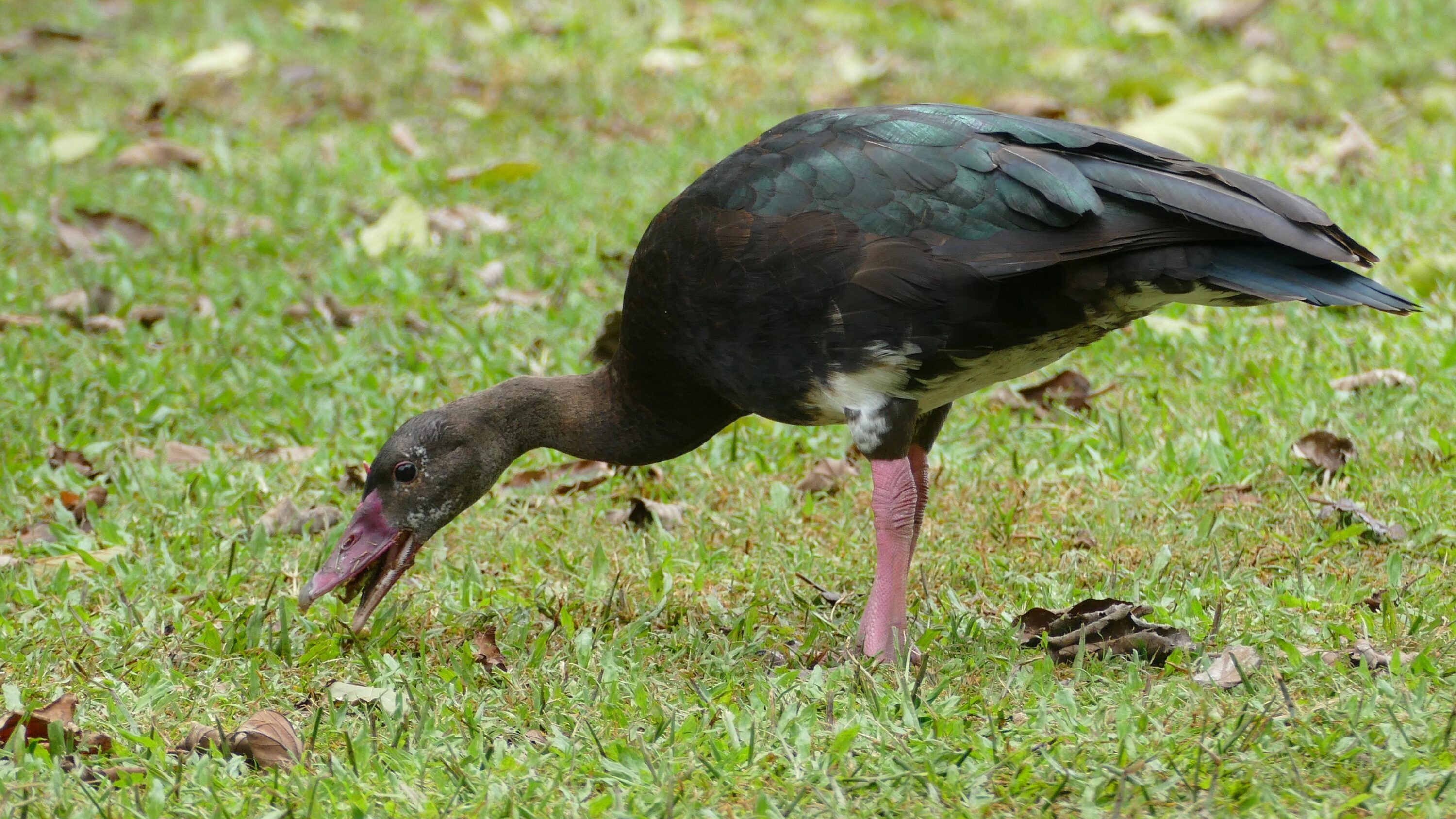
Microscopy Study Described the Respiratory System of Adult Spur-winged Geese
A 2026 microscopy study of ten adult spur-winged geese in Nigeria described the nasal cavity, larynx, trachea, bronchi…
Read story →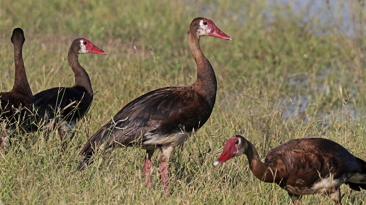
Anatomical Study Documented Feeding Structures and Sex Differences in the Spur-winged Goose Digestive Tract
A 2026 study of ten adult spur-winged geese from Jere, Borno State, Nigeria, described the beak, tongue, oesophagus…
Read story →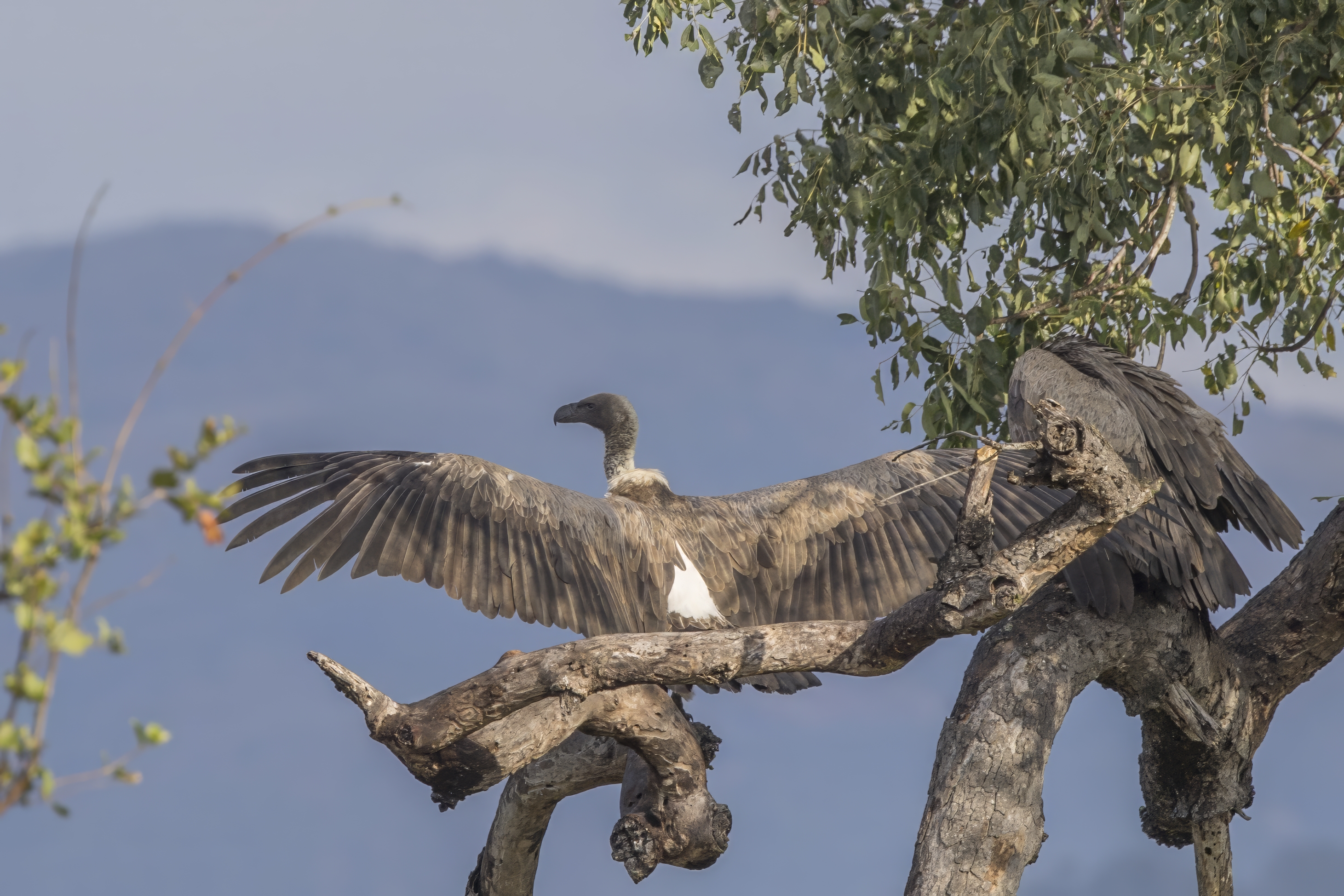
CT Reveals the Organs and Blood Vessels Inside a White-backed Vulture
A contrast-enhanced CT scan of one adult male white-backed vulture revealed major organs and vessels, while also showing…
Read story →SPECIES IN THIS STORY
Species in this story

Independent research and conservation news archive
Did you find this information useful?
Wildlife Vagabond is independently built and maintained. Voluntary support helps cover source verification, hosting and continued work on research and conservation news.
Support Wildlife VagabondThe news archive will remain freely available.