
Great Egret Hearts Revealed Elastic Cartilage in the Pulmonary-Valve Support
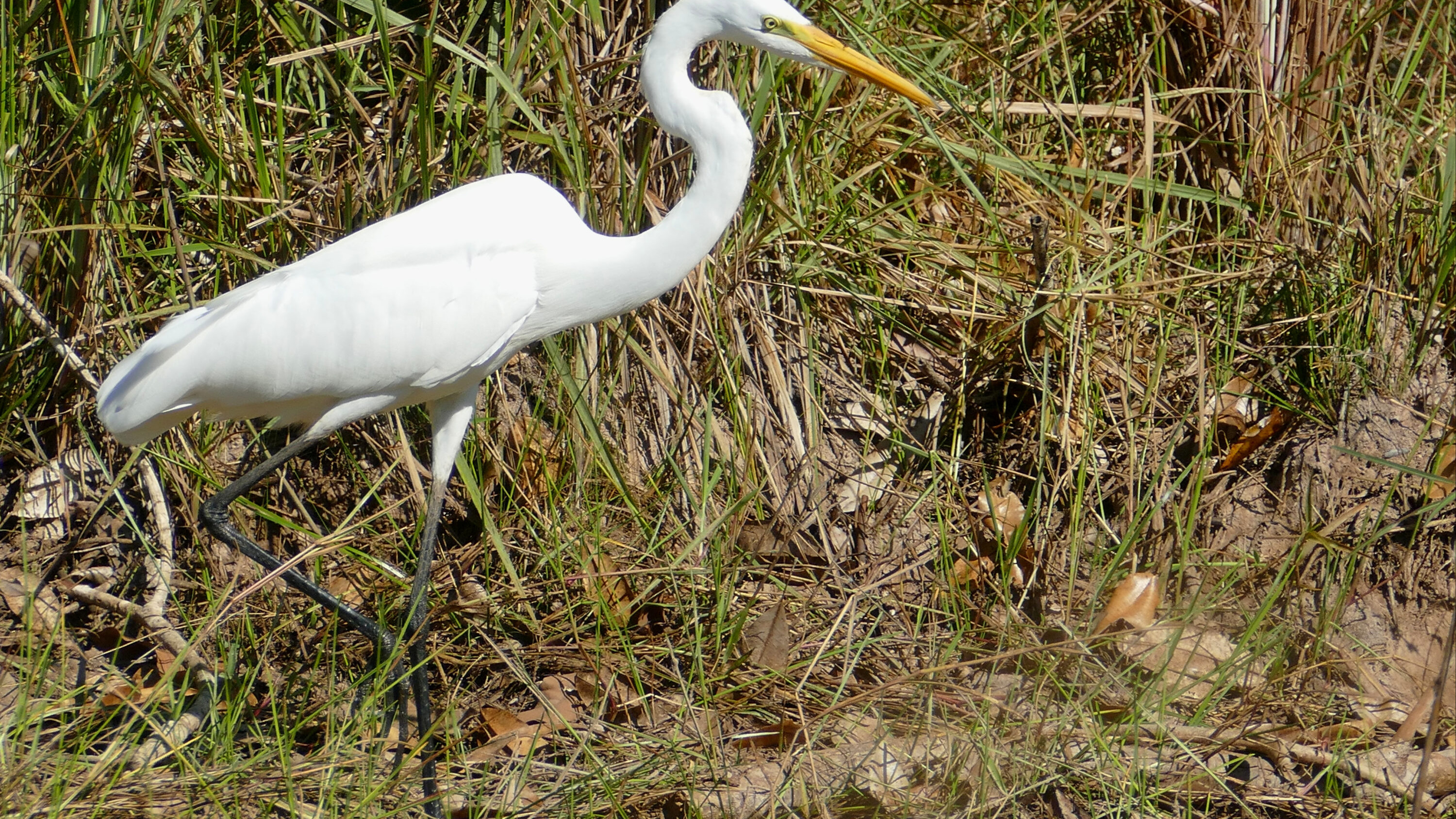
An anatomical study of the heart of great egret (Ardea alba) found elastic cartilage in the pulmonary-valve region of most histologically examined birds — a type of cardiac cartilage the authors say has not previously been reported in vertebrates.
The study, published on 19 May 2026, examined ten adult great egrets from Belém in northern Brazil using radiography, anatomical measurements, histology, scanning electron microscopy and three-dimensional scanning. Eight of the hearts were used for the detailed microscopic cartilage analysis.
The cardiac skeleton helps support the valves and vessel openings
The heart contained the main structural features expected in birds, including four chambers, a thick-walled left ventricle and Purkinje fibres beneath the inner heart lining. Purkinje fibres are specialised cardiac muscle cells that rapidly conduct electrical impulses through the ventricles and help coordinate contraction.
The researchers also described the cardiac skeleton — a framework of dense connective tissue around the bases of the major vessels and valves. This framework included a fibrous ring and a fibrous trigone. Such structures help stabilise the openings of the heart where tissue is repeatedly exposed to mechanical tension.
Two different types of cartilage occurred in the supporting framework
Hyaline cartilage was present in the fibrous trigone and in the commissures of the aortic valve in all eight hearts examined histologically. A commissure is the region where adjacent valve leaflets meet and attach, so cartilage there forms part of the valve’s supporting structure.
The most unusual finding concerned the pulmonary valve, which controls blood flow from the right ventricle into the pulmonary artery. Elastic cartilage was identified in its commissures in six of the eight hearts. The remaining two had hyaline cartilage in the same region instead.
Hyaline cartilage is dominated by collagen and provides firm structural support. Elastic cartilage also contains a dense network of elastic fibres, making it more flexible and able to return to shape after deformation. The study demonstrated the tissue type, but did not test whether this difference changes valve mechanics in living birds.
The function and wider distribution remain unknown
The authors describe the elastic cartilage as the first reported occurrence of this cartilage type in a cardiac cartilaginous structure. They suggest that its presence may be related to the naturally high abundance of elastic fibres in valve tissue, but this remains a morphological interpretation rather than a tested functional mechanism.
Previous studies have described hyaline cartilage, fibrocartilage or even bone in the cardiac skeleton of several birds and mammals. The great-egret finding therefore expands the known range of tissues that can occur in vertebrate hearts and raises a comparative question: whether elastic cartilage has simply been overlooked in other species or represents a more restricted anatomical specialisation.
Anatomy & morphology
Related news
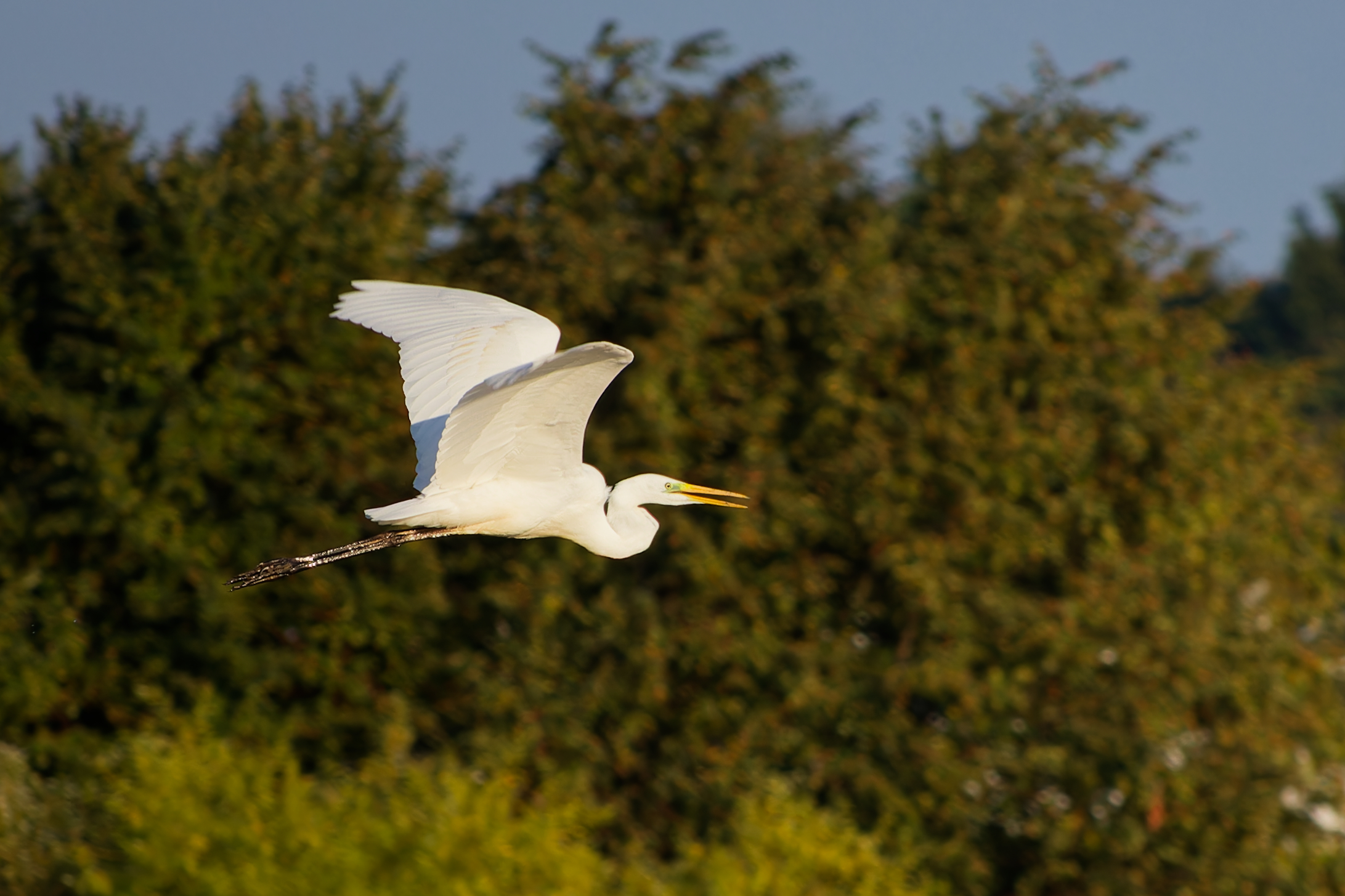
Six of Seven GPS-Tracked Great Egrets Returned to the Same Wintering Grounds
Six of seven great egrets tracked across at least two complete winters returned to the same wintering grounds;…
Read story →
Poland’s Breeding Great Egret Population Reached 1,150–1,250 Pairs
Poland’s breeding great egret population increased by 84% from 2013 to 2025. Breeding was documented at 31 sites,…
Read story →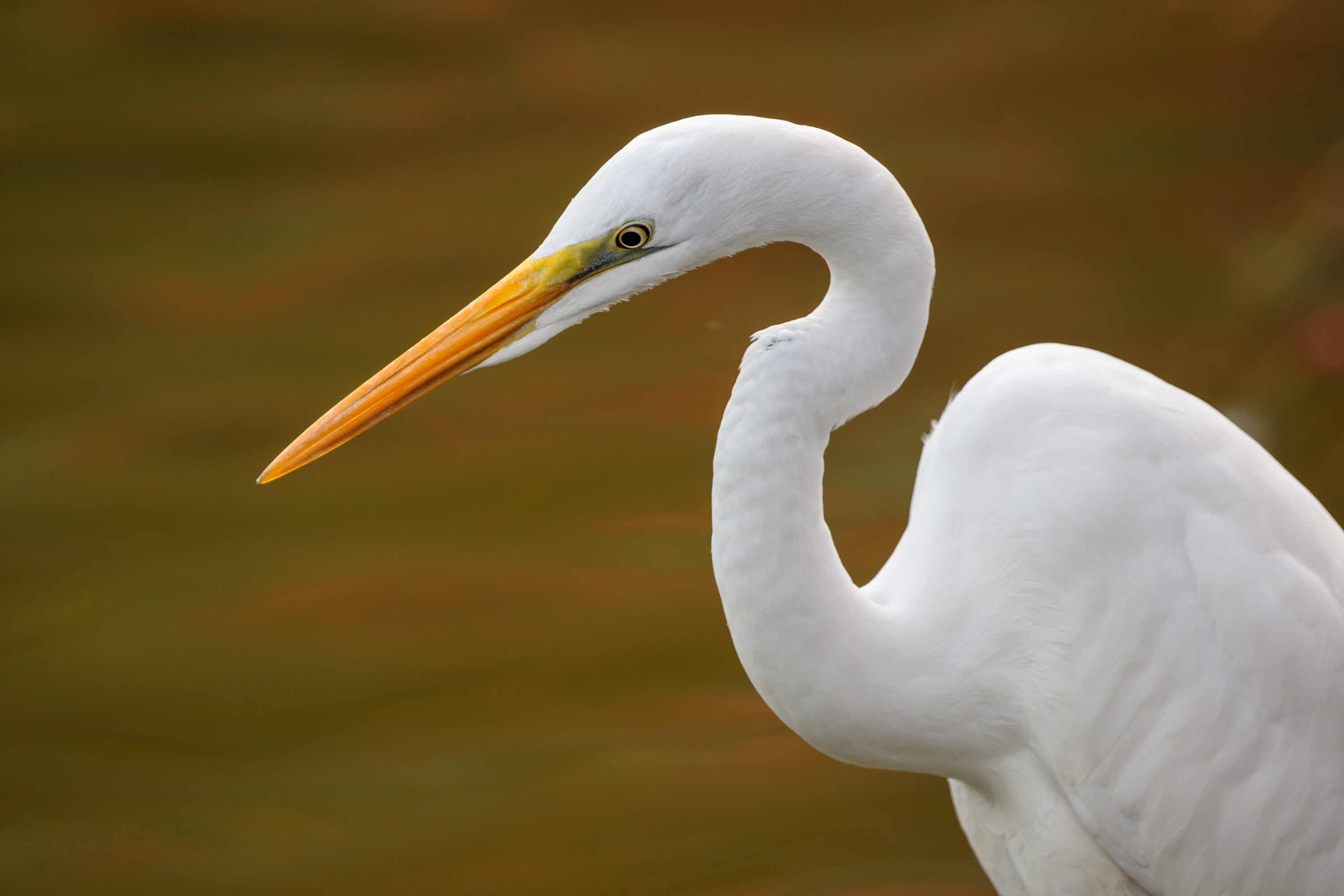
Bile-Duct Trematodes Found in the Lungs and Body Cavity of a Great Egret
A case report from South Korea found the bile-duct trematode Pegosomum bubulcum on the liver surface, in the…
Read story →SPECIES IN THIS STORY
Species in this story
Independent research and conservation news archive
Did you find this information useful?
Wildlife Vagabond is independently built and maintained. Voluntary support helps cover source verification, hosting and continued work on research and conservation news.
Support Wildlife VagabondThe news archive will remain freely available.