
Common Kestrel Retina Had Two Deep, Cone-Dominated Foveal Pits
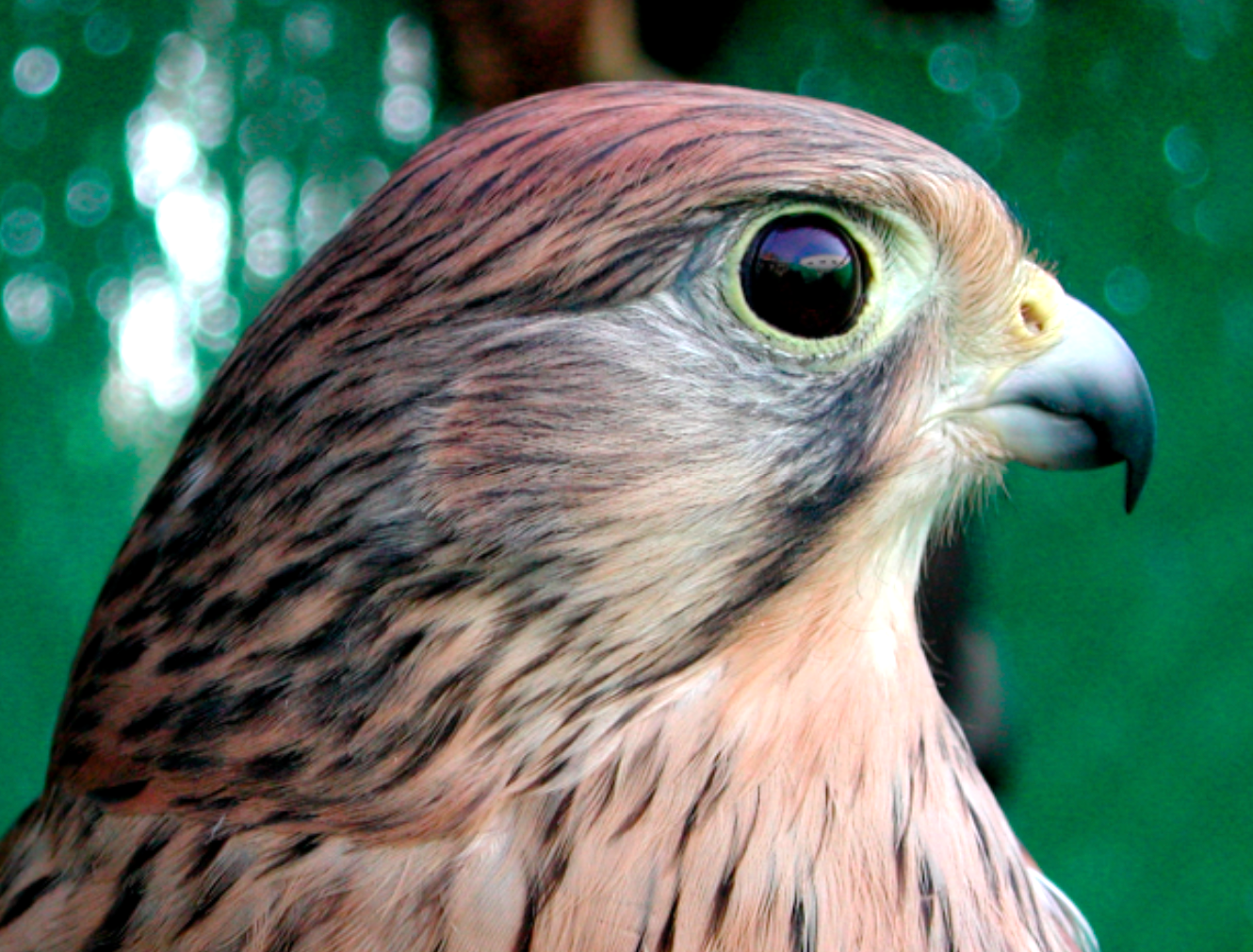
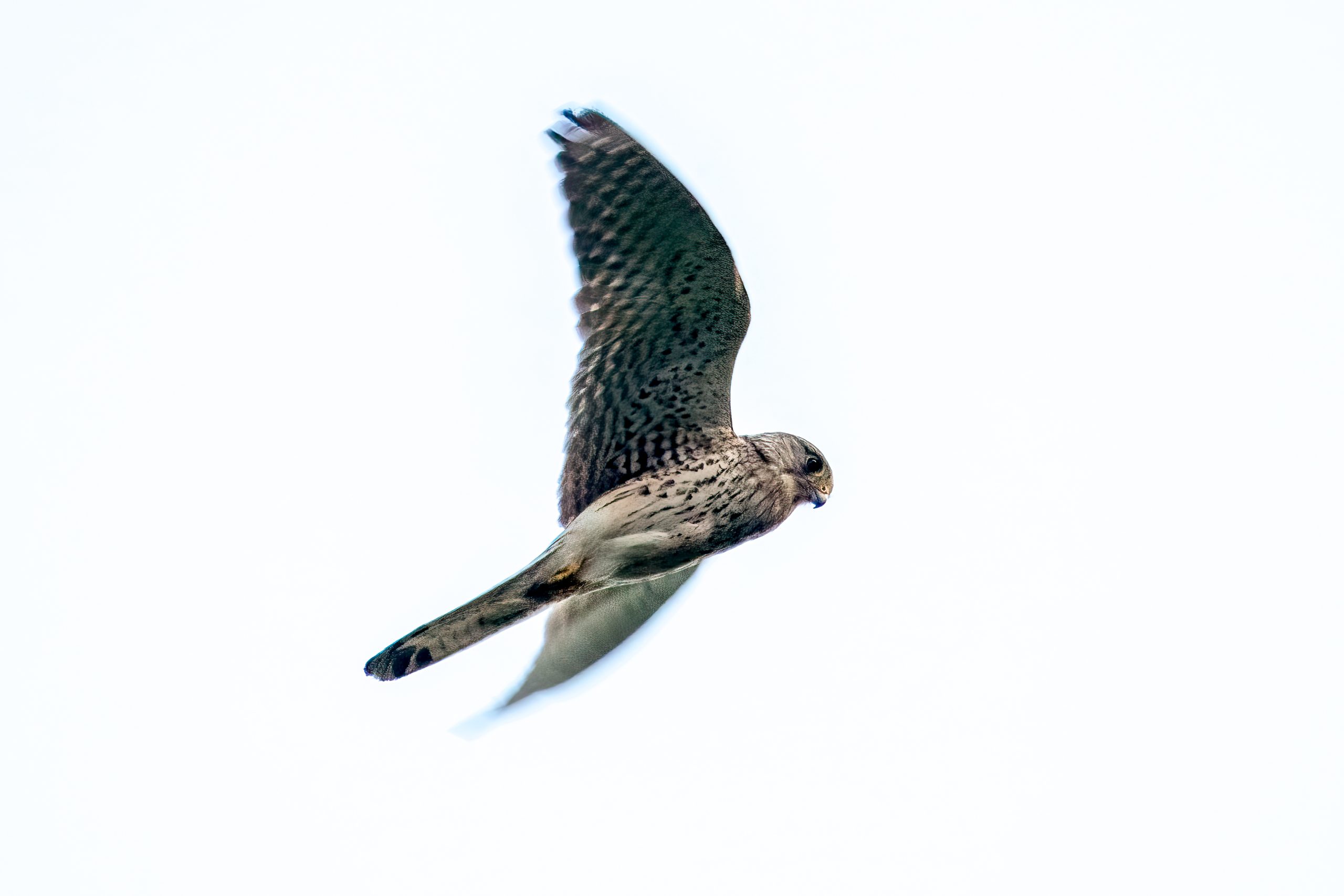
A detailed anatomical study of the common kestrel (Falco tinnunculus) found two distinct deep foveae in the retina and marked structural differences from the centre to the periphery. A fovea is a specialised retinal depression whose structure is typically associated with very high spatial resolution. The work was published on 17 June 2026 in Biology.
The researchers examined the eyes of two adult male kestrels that became available through the Santa Faz Wildlife Recovery Centre after severe injuries unrelated to the eyes made rehabilitation and release impossible. The birds were euthanised on veterinary grounds rather than for the study, and no gross ocular abnormalities were found. The team measured the eyes, prepared histological sections of different retinal regions and used immunolabelling to examine photoreceptor opsins in the foveae.
The second fovea was also unusually deep
The central retina was the thickest region measured, at about 254 µm compared with about 109 µm in the peripheral retina. Two specialised foveae were identified. The central fovea lay within the area centralis and was about 218 µm deep, while the temporal fovea was about 106 µm deep and also had steep walls. In many bifoveate raptors the second, temporal fovea is shallower, so the pronounced temporal pit is a notable feature of the kestrel retina. Both foveae showed centrifugal displacement of the inner retinal layers around the pit, which reduces tissue in the path of incoming light and is characteristic of retinal regions specialised for high spatial resolution.
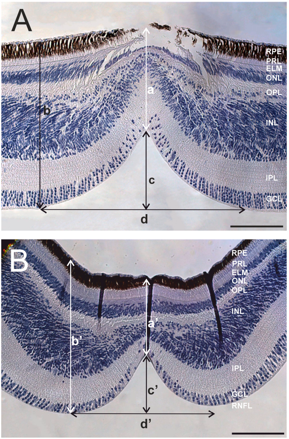
Both foveal pits were cone-dominated
Both foveae showed immunoreactivity for S- and L/M-opsins, visual pigments associated with cone photoreceptors that support colour vision and bright-light vision. Rhodopsin, the visual pigment characteristic of rods used especially in dim light, was not detected within the pit of either fovea, although rhodopsin-positive cells reappeared outside the pits. The authors therefore interpret both foveal pits as predominantly cone-dominated regions consistent with high-acuity daylight vision.
The study did not measure visual acuity behaviourally, nor did it directly count cone or ganglion-cell density. The functional interpretation rests on the anatomy of the deep foveae, displacement of inner retinal layers and the photoreceptor markers detected there.
The study provides a detailed morphological and histological baseline for the kestrel visual system. The authors note that only two specimens were available, but the results add species-specific information on how the bifoveate retina of a diurnal falcon is organised.
Anatomy & morphology
Related news
Kestrels Had More Densely Packed Mucous Glands Than Storks
A small study in Türkiye found roughly twice the density of mucus-producing glands in common kestrels’ windpipes as…
Read story →
No Common Kestrels Were Recorded at Rani for Three Consecutive Winters
Fourteen winters of surveys at Rani in Assam produced 422 common kestrel records, but sightings declined after 2020…
Read story →
Winter Surveys Estimated 5.59 Common Kestrels and 2.61 Common Buzzards per km² in Parma Farmland
Roadside distance sampling in northern Italy estimated 5.59 common kestrels and 2.61 common buzzards per km², while the…
Read story →SPECIES IN THIS STORY
Species in this story
Independent research and conservation news archive
Did you find this information useful?
Wildlife Vagabond is independently built and maintained. Voluntary support helps cover source verification, hosting and continued work on research and conservation news.
Support Wildlife VagabondThe news archive will remain freely available.