
European Robin Retina Study Mapped Transducin Subunits and Found No Gtγ10 in Photoreceptors

A molecular study of the European robin (Erithacus rubecula) has mapped several components of transducin, a G-protein complex that helps photoreceptor cells convert light into a cellular signal. The work is particularly relevant because European robins use a light-dependent magnetic compass during migration, and some transducin subunits have been proposed as possible partners of cryptochrome 4a (Cry4a), a blue-light receptor involved in a leading hypothesis for magnetic sensing.
Mapping the signalling machinery of rods and cones
The researchers examined retinal tissue from adult robins captured near the University of Oldenburg in Germany. They combined single-cell RNA sequencing, which shows which genes are active in individual retinal cell types, with immunohistochemistry to localise the corresponding proteins in retinal tissue. Separate biochemical assays tested whether selected transducin β- and γ-subunits were physically capable of forming complexes.
Transducin consists of α, β and γ subunits. Different combinations are used in rods and cones, the two major classes of light-sensitive cells in the vertebrate retina. In the robin RNA data, GNB1, which encodes Gtβ1, was associated mainly with rods, while GNB3, encoding Gtβ3, was the principal β-subunit in cones. Among the γ-subunits, GNGT2, encoding GtγT2, was dominant in cones, whereas GNG11, encoding Gtγ11, was associated most strongly with rods.
The protein staining broadly supported this division but also showed that the picture is not perfectly simple. GtγT2 protein was detected in both rods and cones, while Gtγ11 stained rods strongly and red and double cones more weakly. Gtβ3 staining was broader than the RNA result suggested; the authors note that some of this signal could reflect cross-reactivity of the antibody with the closely related Gtβ1 protein.
Gtγ10 was missing from the photoreceptors
The clearest negative result concerned Gtγ10. Earlier work had identified GNG10, the gene encoding this subunit, as a possible Cry4a interaction partner, making it a candidate for a molecular link between light signalling and magnetoreception. In the robin study, however, GNG10 expression was not detected in rods or cones, and an antibody against Gtγ10 likewise produced no specific signal in either photoreceptor type.
Gtγ10 was not absent from the retina altogether. The transcript data placed GNG10 in other retinal cell classes, including retinal ganglion cells, oligodendrocytes and Müller glia. The result therefore argues specifically against a role for Gtγ10 in the photoreceptors themselves rather than showing that the molecule is absent from the eye.
Laboratory binding did not mean the proteins meet in the retina
The biochemical interaction experiments produced an apparently different result. Gtβ3 was able to form βγ complexes with all three γ-subunits tested — GtγT2, Gtγ10 and Gtγ11 — in pulldown, co-immunoprecipitation and NanoBiT assays. This shows that the proteins are chemically capable of associating under the experimental conditions.
But the authors argue that physical compatibility alone is not enough to determine which complexes actually form inside a robin photoreceptor. A protein pair can only interact in vivo if both components are expressed in the same cell. Because Gtγ10 was not detected in rods or cones, the in-vitro ability of Gtβ3 and Gtγ10 to bind does not make that pair a likely photoreceptor complex. The cell-specific expression pattern instead supports Gtβ3/GtγT2 as a likely cone combination and Gtβ1 with rod-associated γ-subunits in rods.
A narrower candidate for magnetoreception
The study also found that Cry4 and Gtα2, another proposed Cry4a interaction partner, co-localised in photoreceptor outer segments likely belonging to red cones and double cones. This agrees with earlier work suggesting that these cells may be relevant to the robin’s magnetic compass. The antibody used for Cry4 could not distinguish the Cry4a and Cry4b isoforms, however, so the staining does not by itself demonstrate a specific Cry4a–Gtα2 complex.
Taken together, the results do not support Gtγ10 as a photoreceptor component of a Cry4-based magnetoreception pathway. The authors suggest that if a γ-subunit of transducin participates in such a mechanism, GtγT2 is a more plausible candidate because it is actually present in cone photoreceptors. This remains a molecular inference rather than a demonstration of magnetic sensing: the study did not manipulate the birds’ magnetic orientation or show that GtγT2 is required for compass behaviour.
The work is presented as the first study of G-protein subunit expression in a migratory songbird. It was first posted as a bioRxiv preprint on 29 June 2026 and was subsequently revised. As a preprint, it had not undergone journal peer review at the time of the latest version.
Molecular biology & biochemistry
Related news

Shorter Days Altered Migratory Restlessness in Juvenile Robins but Not Garden Warblers
In a Lund experiment, juvenile robins exposed to a southern-France photoperiod showed a weaker rise in nocturnal migratory…
Read story →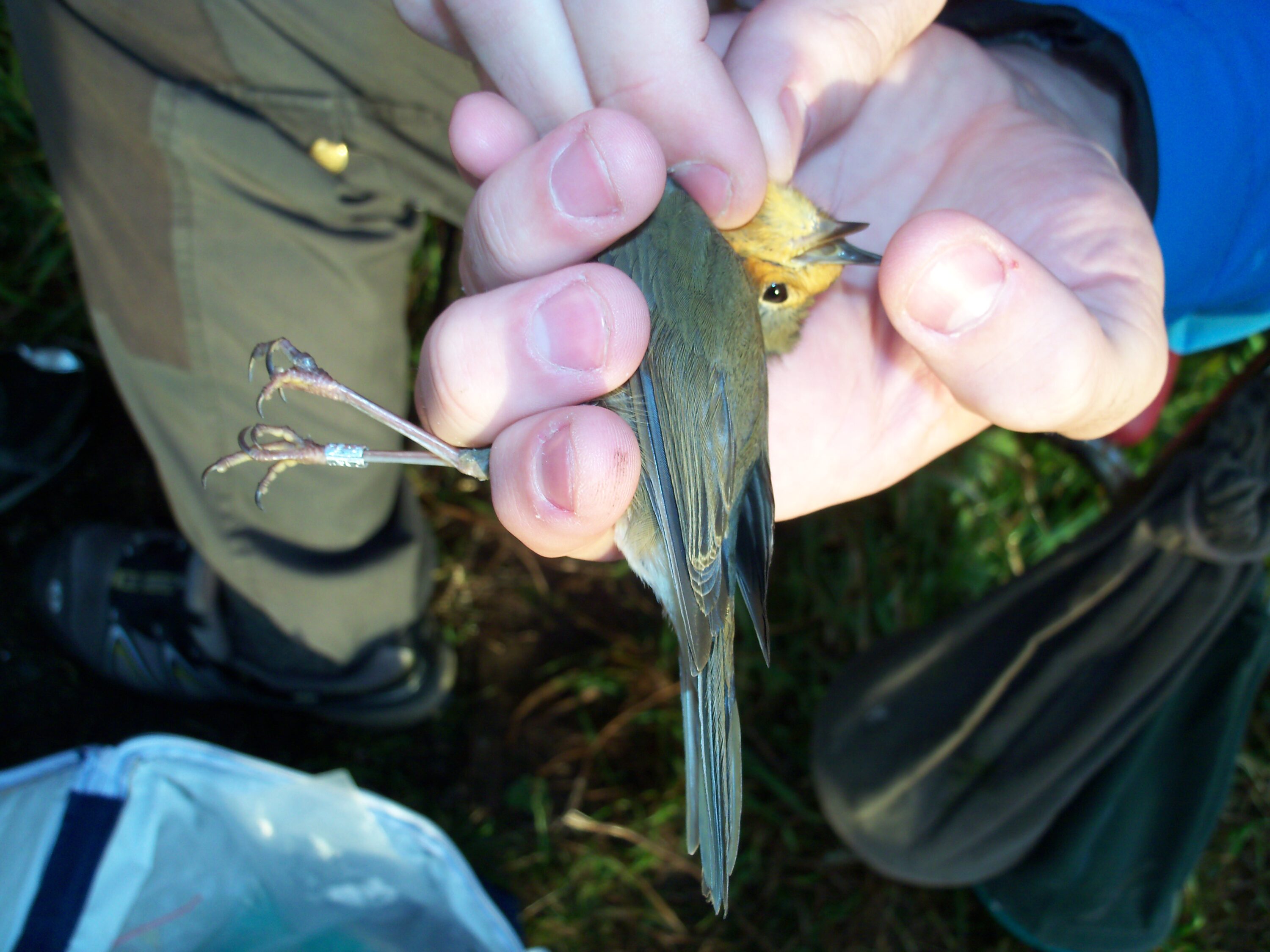
Early-Migrating Robins Carried More Ticks Than Late Migrants
Nearly half of 198 migrating European robins carried ticks, and early autumn migrants had significantly higher tick loads…
Read story →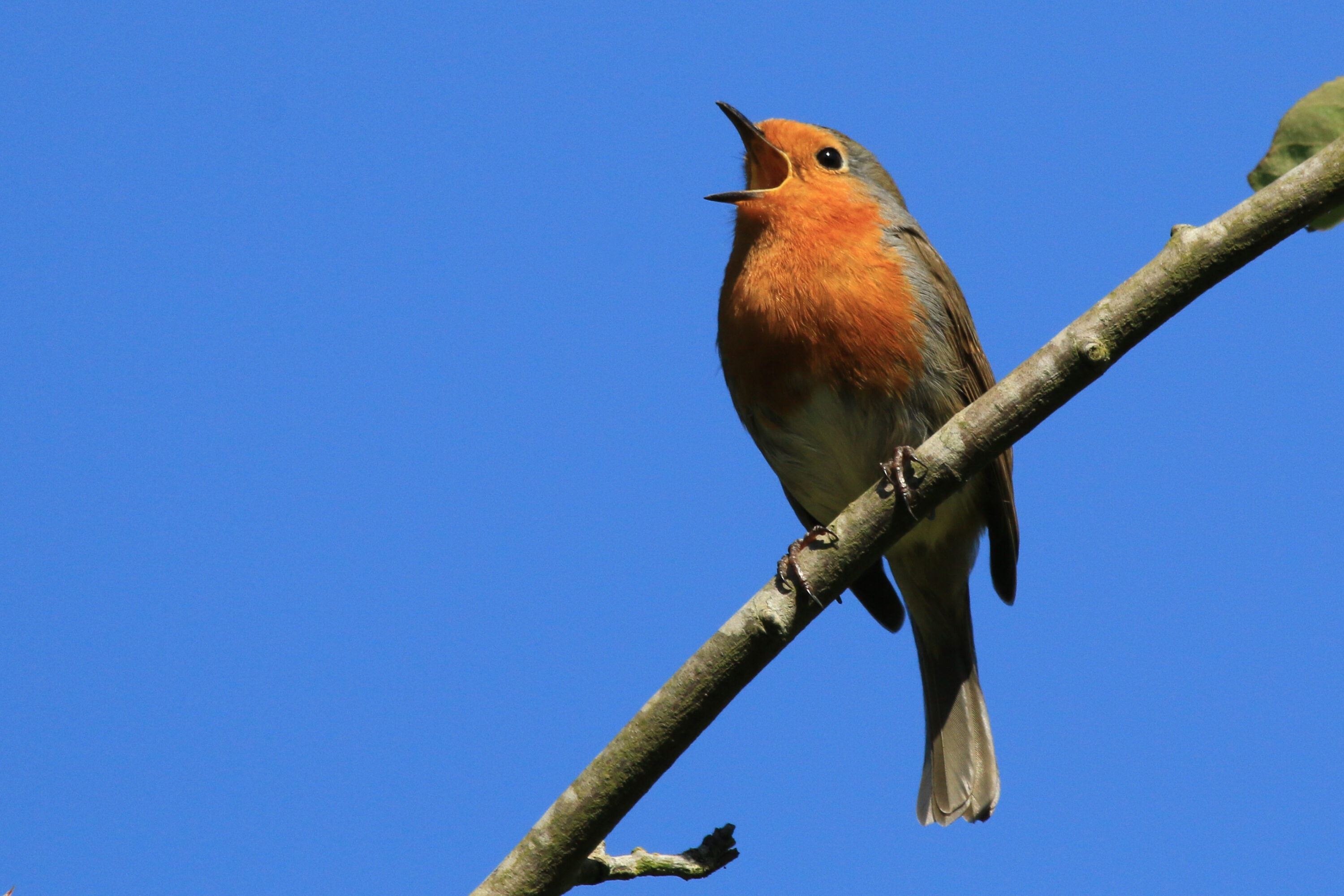
Site-Specific Thresholds Improved Robin Detection Precision by 15–29 Percentage Points
A 2026 protocol using European robin recordings improved automated detection precision by about 15–29 percentage points across three…
Read story →SPECIES IN THIS STORY
Species in this story

Independent research and conservation news archive
Did you find this information useful?
Wildlife Vagabond is independently built and maintained. Voluntary support helps cover source verification, hosting and continued work on research and conservation news.
Support Wildlife VagabondThe news archive will remain freely available.