
How one rhesus macaque’s brain represented coordinated finger movements
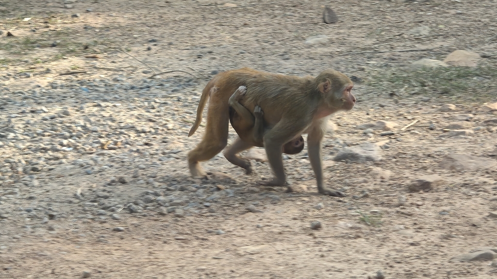
When two fingers move together, does the brain simply combine the activity patterns it uses to move each finger on its own? Recordings from one rhesus macaque suggest the answer is no. In three connected cortical areas involved in grasping, activity during coordinated finger movements could not be fully explained by simply adding together the patterns seen during single-finger movements.
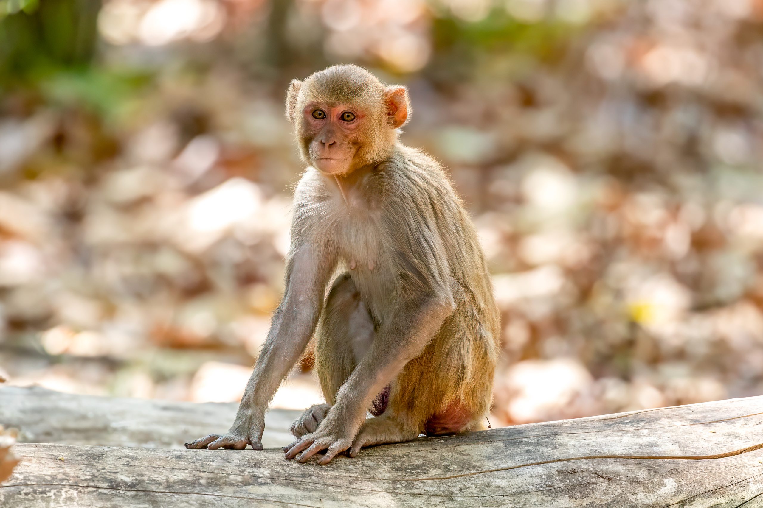
Researchers recorded activity from groups of neurons in the anterior intraparietal area (AIP), ventral premotor area F5 and primary motor cortex (M1) while a male Macaca mulatta flexed individual fingers or pressed two fingers together using a microswitch device. The task included single-finger movements and two-finger combinations. These brain areas form part of the macaque grasping network. By comparing their activity across the instructed movements, the researchers could ask how differently each area represented the movements.
Coordinated movements had their own activity patterns
All three areas showed different activity patterns for the five instructed movements. But the patterns for combined movements were not just straightforward combinations of the patterns for their component fingers. For example, activity during a combined index- and middle-finger movement was positively correlated with the pattern for middle-finger movement and negatively correlated with the pattern for index-finger movement. This points to a representation more complicated than a simple average of the two single-finger patterns.
The areas also differed in how many independent patterns were needed to describe the movements. For 90% of the movement-related variation, F5 required an average of 17.8 independent patterns, compared with 11.8 in AIP and 11.7 in M1. In other words, activity recorded in F5 varied across the instructed movements in more independent ways. This difference was supported across recording sessions. It does not mean that F5 simply controlled more fingers; it shows that movement-related activity there had a more complex pattern of variation across the task.
Brain activity during mistakes differed across areas
On error trials, when the macaque performed a movement other than the one instructed, activity in AIP and F5 initially matched the instructed movement more closely than the movement actually made. As the trial progressed, AIP remained more instruction-related, M1 shifted toward the movement the animal performed, and F5 showed an intermediate pattern. By the time of the erroneous press, M1 activity preferentially matched the movement actually produced. This relative ordering across areas is descriptive: the study did not directly test the differences between areas statistically.
Intracortical brain-computer interfaces use signals recorded from inside the brain to control an external device. A decoder that learns to interpret signals during single-finger movements might not interpret combined-finger movements equally well, and the results suggest that this could depend partly on which brain area supplies the signals. That possibility was not tested directly: the study did not compare decoder performance, and it involved one macaque. Its contribution is to show that coordinated finger movements are represented in distinct, area-dependent ways—an important consideration for future tests of how brain signals can be translated into movement commands.
Physiology
Related news
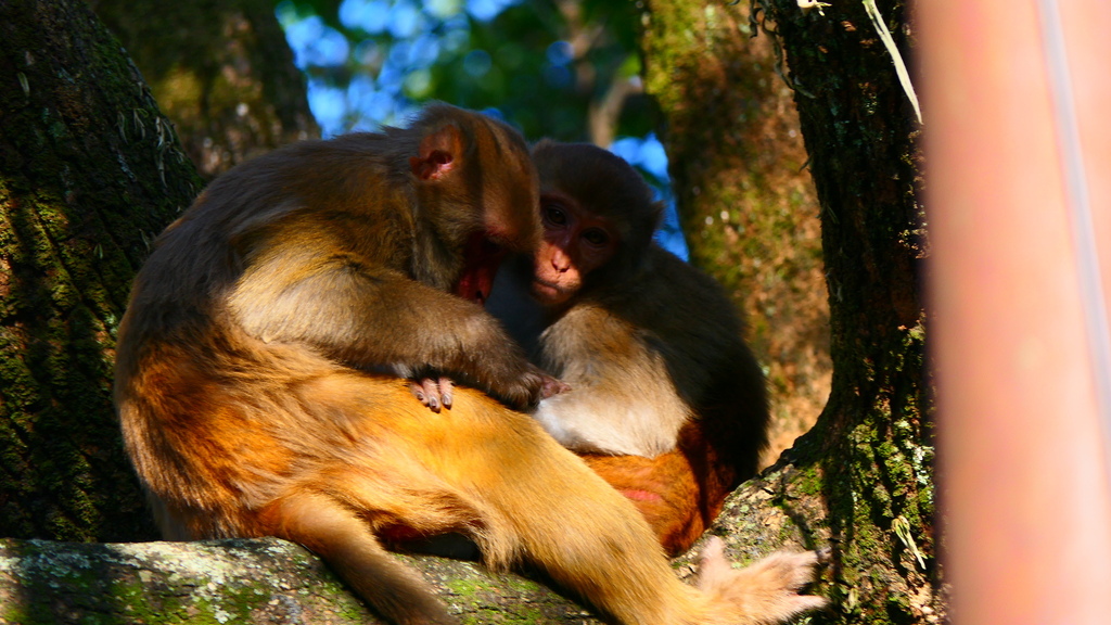
A macaque antibody neutralised diverse H5 flu viruses by blocking membrane fusion
An antibody isolated from immunised rhesus macaques neutralised H5 influenza viruses collected from 1997 to 2024, including recent…
Read story →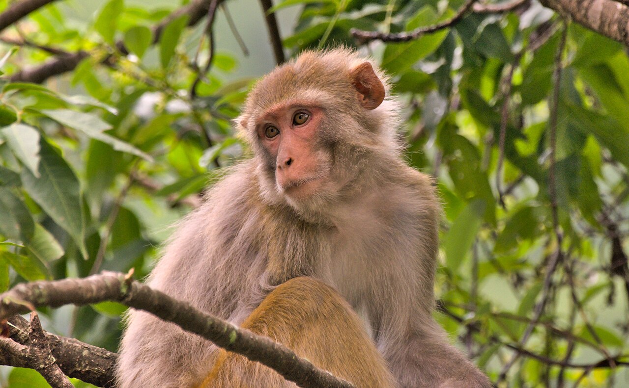
Female Rhesus Macaques That Watched Unfamiliar Female Faces Longer Were More Likely to Have Offspring the Following Year
At a semi-free-ranging population in Puerto Rico, females that spent longer looking at an unfamiliar female’s neutral face…
Read story →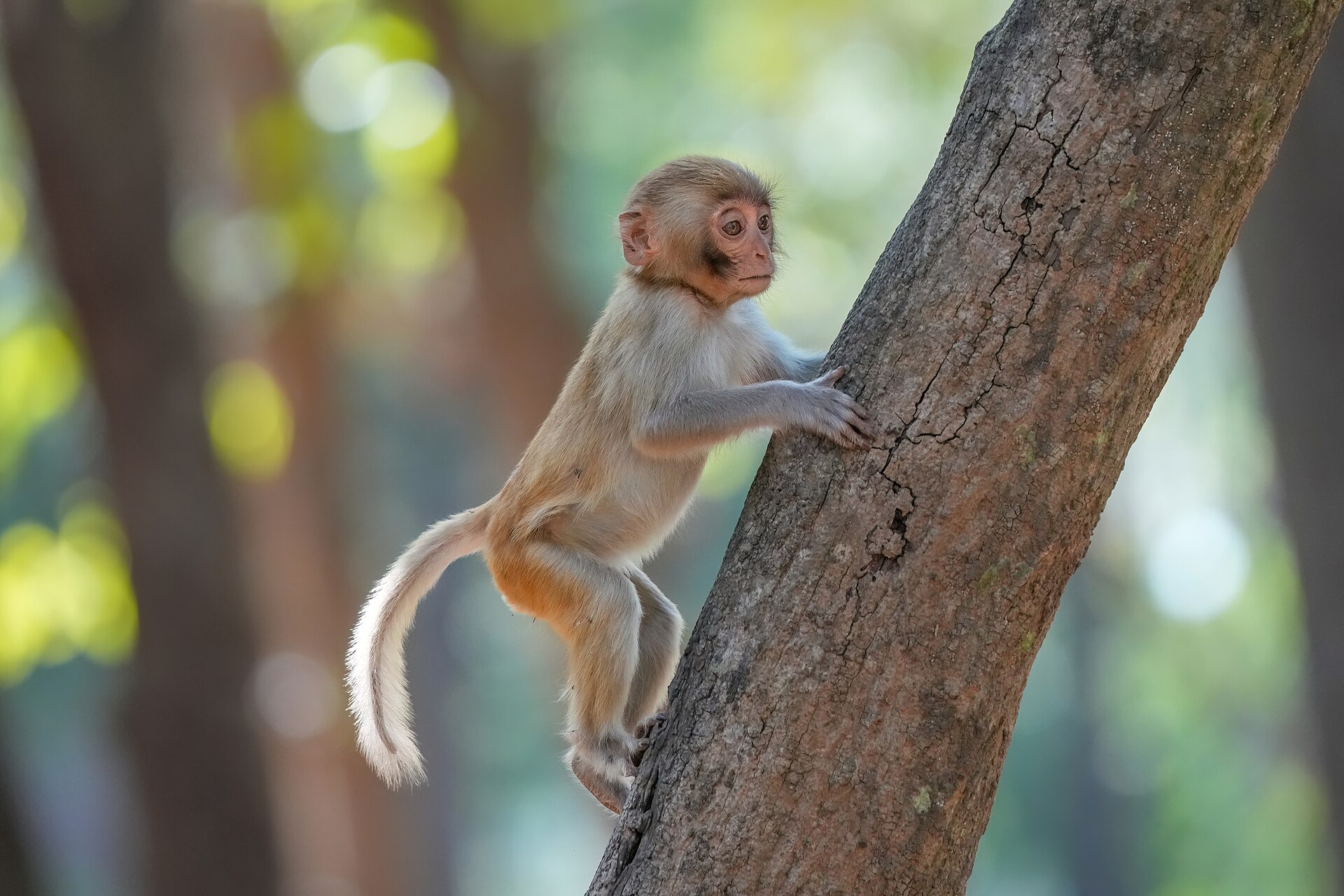
Rhesus macaques slow down after errors, but do not perform better
In tasks answered with eye movements or a touchscreen, three rhesus macaques took longer to respond after making…
Read story →SPECIES IN THIS STORY
Species in this story
Independent research and conservation news archive
Did you find this information useful?
Wildlife Vagabond is independently built and maintained. Voluntary support helps cover source verification, hosting and continued work on research and conservation news.
Support Wildlife VagabondThe news archive will remain freely available.