
Pigeon Neural Populations Encoded Specific Movements Rather Than Simply Identifying Other Pigeons

Neural populations in the pigeon brain responded to specific features of another pigeon’s movements rather than simply signalling whether a viewed stimulus was a pigeon, according to a study published on 26 February 2026. Researchers recorded individual nerve cells in three brain regions while pigeons (Columba livia) watched videos of other pigeons and control shapes.
The study used 11 pigeons. The head-fixed and body-restrained birds viewed videos showing four kinds of behaviour—courtship, feeding, flight and walking—while researchers recorded single-unit activity in the visual Wulst, the mesopallium ventrolaterale (MVL) and the nidopallium caudolaterale (NCL). These regions contribute to different aspects of visual processing and cognition.
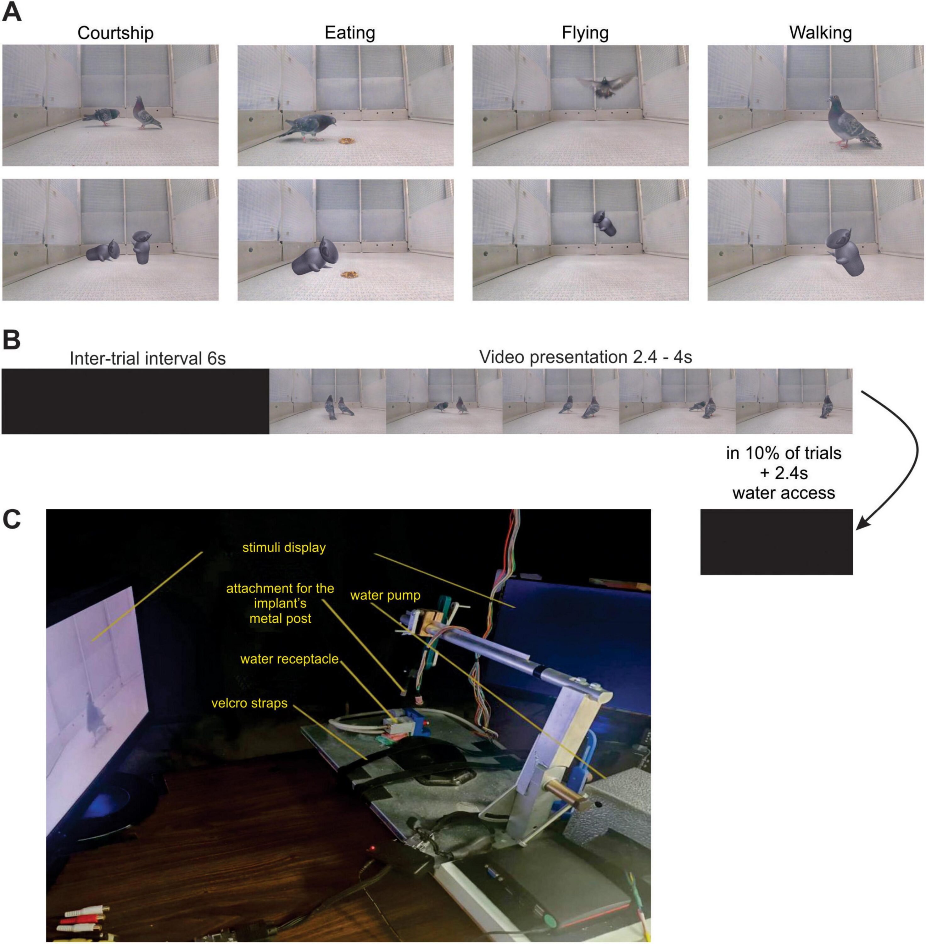
Single neurons did not simply code “pigeon”
Visually responsive neurons were found in all three regions, but the researchers did not find a categorical separation in which individual neurons consistently distinguished videos of pigeons from the control stimuli.
Population-level analyses in the two more directly visual regions, MVL and the visual Wulst, instead revealed brief changes in neural activity that coincided with particular movement features. Responses were associated with bowing, wing-flapping and head-bobbing, indicating that groups of neurons encoded components of the observed behaviour rather than maintaining a simple “conspecific” category signal.
Visual pathways differed in how they combined information
Some visually responsive neurons were also influenced by sound. These sound-modulated visual cells were significantly more common in MVL than in the visual Wulst. The authors interpret this as a sign that visual and auditory information may be combined earlier than previously recognised in the tectofugal pathway, one of the major visual pathways in the bird brain. They also caution that sound-modulated cells made up only a small part of the recorded population, so this result needs further testing.
During passive viewing, NCL did not show the same clear modulation by visual features as the two more directly visual regions. The authors therefore suggest that NCL was less involved in automatic analysis of these visual details under the conditions tested.
Physiology
Related news

Inland Mute Swans Had Higher Winter Body Condition Despite Harsher Winters and Supplemental Feeding
A study of 853 mute swans from four Polish wintering regions found higher body condition inland than on…
Read story →
pH-Adjusted Hill Equation Reliably Estimated Blood Oxygen Saturation From 70–100% in Immobilised White Rhinos
A South African study of 16 immobilised male white rhinos in Kruger National Park found that a pH-adjusted…
Read story →
Task Uncertainty Increased Interference From Irrelevant Visual Information in Rhesus Macaque Decisions
Two rhesus macaques made less accurate visual decisions under task uncertainty; recordings and microstimulation in primary visual cortex…
Read story →SPECIES IN THIS STORY
Species in this story
Independent research and conservation news archive
Did you find this information useful?
Wildlife Vagabond is independently built and maintained. Voluntary support helps cover source verification, hosting and continued work on research and conservation news.
Support Wildlife VagabondThe news archive will remain freely available.