
Fascioloides magna Infection Produced Liver Scarring and Regenerative Nodules in Red Deer
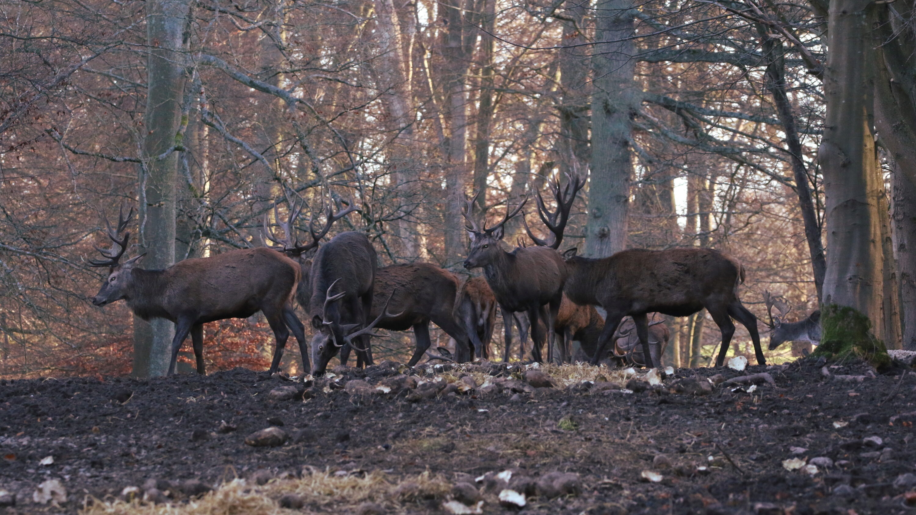
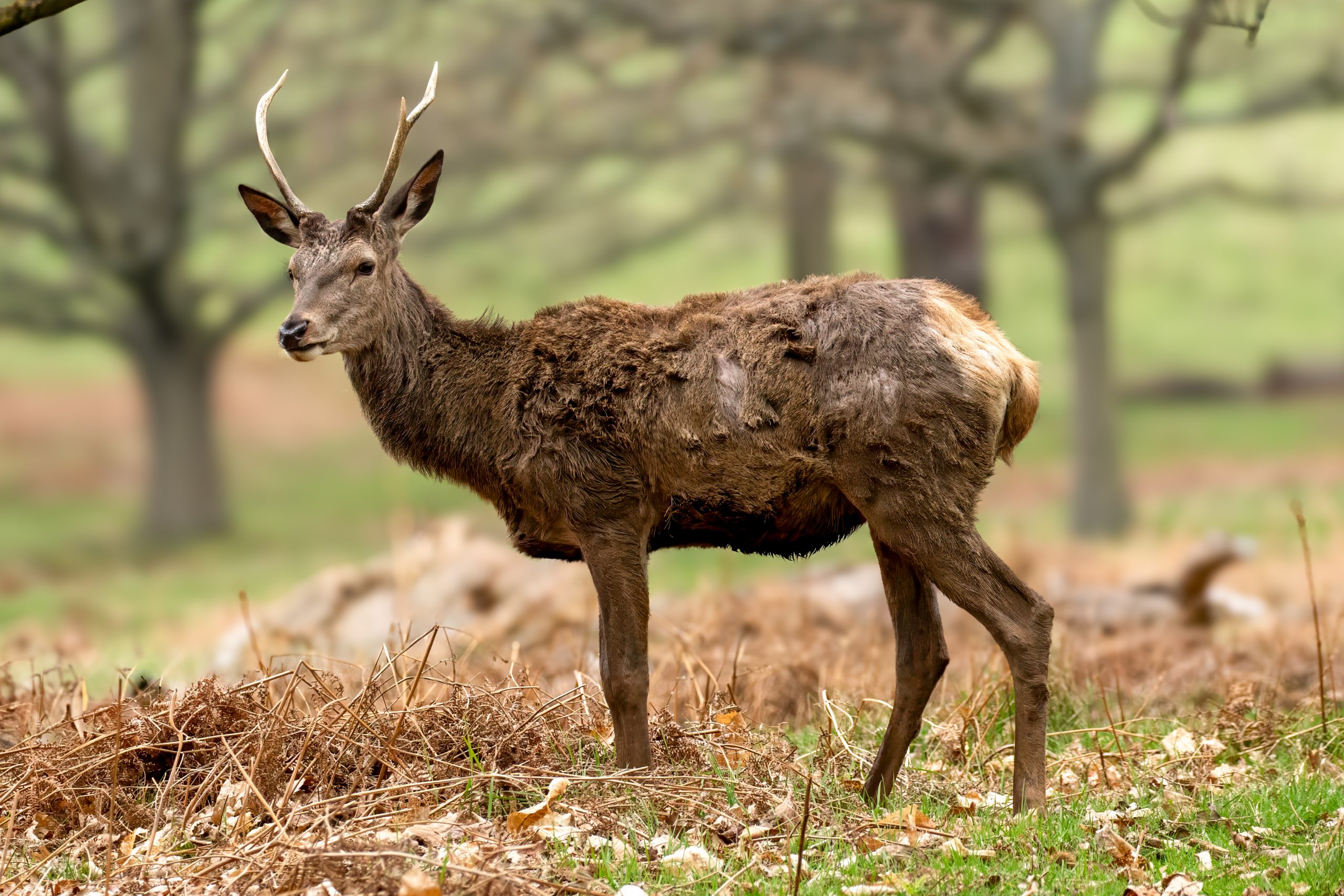
A study of red deer (Cervus elaphus) livers from Croatia documented extensive liver damage caused by the giant liver fluke Fascioloides magna, together with areas where damaged tissue was being partially rebuilt. Researchers examined 136 livers and followed lesions ranging from migrating juvenile flukes and established pseudocysts to degrading pseudocysts after antiparasitic treatment. A pseudocyst is a capsule-like structure formed by the host around adult flukes in the liver; it limits continued parasite migration and tissue damage while allowing the flukes to survive and reproduce.
Some infected livers contained dozens of flukes and pseudocysts
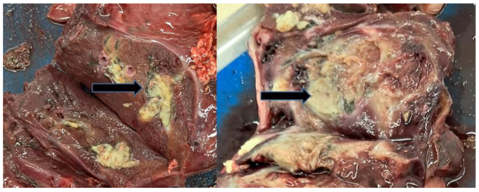
Some infected livers carried a heavy parasite burden, with up to 45 pseudocysts and 70 adult flukes in a single liver. Juvenile flukes were also found migrating through the liver in some animals. The infection produced migratory channels, black iron-porphyrin pigment, inflammation and fibrous tissue, while treated pseudocysts could contain degrading material and remnants of dying flukes.
Repair produced regenerative nodules as well as scar tissue
Some livers contained nodules up to 7 cm across. Microscopic examination showed that these areas were divided by bands of fibrous connective tissue and contained pigment and remnants of trematode eggs. Within the nodules, the researchers found disorganised proliferating hepatocytes — liver cells multiplying during repair — together with fibrous bands and occasional blood vessels.
The sample was not suitable for estimating infection prevalence: only 136 of roughly 600 red deer livers from the county were submitted for examination, and they were obtained through non-random convenience sampling.
The authors describe the response as substantial repair of damaged liver tissue rather than complete restoration of the original structure. Part of the affected area may develop into new hepatic tissue, while the remainder becomes scar tissue. They propose that this response to severe acute and chronic injury could provide a model for studying abnormal liver repair.
Disease, parasites & microbiology
Related news
Velvet Antler Had the Most Distinct Metabolite Profile in Red and Sika Deer
A Chinese study detected 2,361 metabolites across three antler tissues from red and sika deer. Velvet antler separated…
Read story →
Tarim Red Deer Microbiomes Showed Greater Capacity to Break Down Reed Fibre
Under the same reed-containing diet, captive Tarim red deer had more fibre-associated gut microbes, higher cellulase and hemicellulase…
Read story →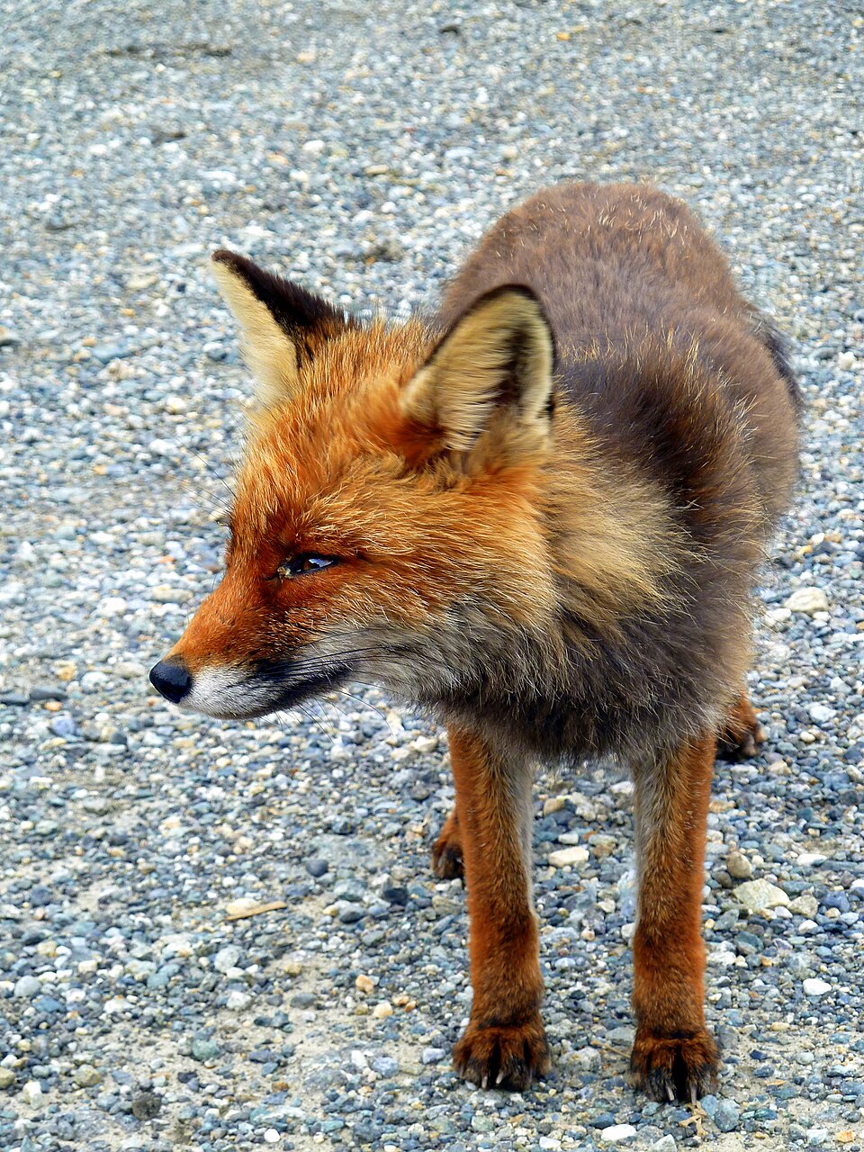
Wolf Kills Provided Plenty of Food for Red Foxes
Red foxes visited all 26 wolf-killed red deer monitored in Stelvio National Park. The amount of meat left…
Read story →SPECIES IN THIS STORY
Species in this story
Independent research and conservation news archive
Did you find this information useful?
Wildlife Vagabond is independently built and maintained. Voluntary support helps cover source verification, hosting and continued work on research and conservation news.
Support Wildlife VagabondThe news archive will remain freely available.