
Aging rhesus macaques show broader protein changes in blood than in brain fluid

As animals age, changes in the brain may also be reflected in the fluids around it and in the bloodstream. To investigate how these patterns differ, researchers measured proteins in blood plasma and cerebrospinal fluid from 66 rhesus macaques (Macaca mulatta) aged about 6–34 years. Cerebrospinal fluid is the fluid that surrounds the brain and spinal cord.
The macaques lived at UC Davis. Researchers measured proteins linked to nervous-system aging, inflammation and neurodegeneration in blood and cerebrospinal fluid, and examined brain tissue from 41 macaques for amyloid and tau, two proteins whose accumulation or altered forms are important in human neurodegenerative disease.
More age-related differences in plasma
Age was associated with differences in about 38% of the measured plasma proteins, compared with about 19% of those measured in cerebrospinal fluid. The plasma differences were mostly increases. Several proteins linked to nerve-cell axons and supporting glial cells were higher in older macaques. Neurofilaments help maintain the internal structure of axons and are widely studied as markers of axonal injury. GFAP is a structural protein in astrocytes, the glial cells that help maintain the environment around neurons. Their higher blood concentrations are therefore relevant to age-related changes in nervous tissue, although the measurements do not establish the cause.
Age-related differences in CHIT1 and TREM2, proteins associated with the brain’s immune cells called microglia, were more apparent in cerebrospinal fluid. TREM2 is involved in microglial responses to changes in nervous tissue. In contrast, neuronal pentraxins (NPTX1, NPTX2 and NPTXR), proteins associated with the organization and function of synaptic connections, were lower in older animals. Pleiotrophin, which is associated with neuronal growth and support, was also lower. These findings suggest that immune-cell and synaptic protein patterns do not change in the same way with age.
Two particularly informative proteins were FOLR1, a receptor that helps transport folate into cerebrospinal fluid, and IGFBP7, a protein linked to cellular senescence and enriched in the choroid plexus, which produces cerebrospinal fluid. Both were lower in the pre-geriatric group. Among geriatric macaques, however, researchers identified two different patterns: some had relatively high levels of both FOLR1 and IGFBP7, while others had low levels of both. The high-level profile also contained more of several synapse-related proteins, whereas the low-level profile was relatively enriched in proteins associated with immune activity and blood-vessel regulation. These observed profiles did not simply follow chronological age and are not established biological subtypes.
Brain tissue showed amyloid accumulation, but little tau pathology
In the brain tissue examined, amyloid deposits accumulated with age, while pathological tau deposits were scarce. This resembles one part of the changes seen in Alzheimer’s disease, where both amyloid and abnormal tau can accumulate, but it does not mean that the macaques showed the full disease process. In plasma, amyloid-β and some phosphorylated forms of tau showed age-related differences; corresponding proteins in cerebrospinal fluid were largely stable. Several other plasma proteins were associated with the amount of amyloid in the brain even after accounting for age, while such associations in cerebrospinal fluid were more limited. Blood and brain tissue therefore revealed related but not identical aspects of aging; the correlations do not show that these proteins caused the deposits.
Two additional plasma proteins tended to be higher with age: IL6, a cytokine involved in inflammatory signalling, and GDF15, a protein associated with cellular and metabolic stress. Their different biological roles help distinguish the broad age-related changes in blood from the more selective changes in the fluid surrounding the brain.
The results suggest that aging-related protein patterns are not the same in the bloodstream and the fluid surrounding the brain. Changes were broader in plasma, while cerebrospinal-fluid differences were more selective and included varied profiles among geriatric macaques. Comparing the two fluids may therefore help researchers understand which protein patterns reflect changes across the body and which are more closely associated with the central nervous system.
The study provides a basis for comparing aging biomarkers in non-human primates and people, but it does not establish what causes the changes or when they develop: the animals were assessed at one point in time. The protein test was developed primarily for human samples, and differences in how its antibodies recognize macaque proteins can affect what is detected and how cross-species comparisons are interpreted.
Physiology
Related news
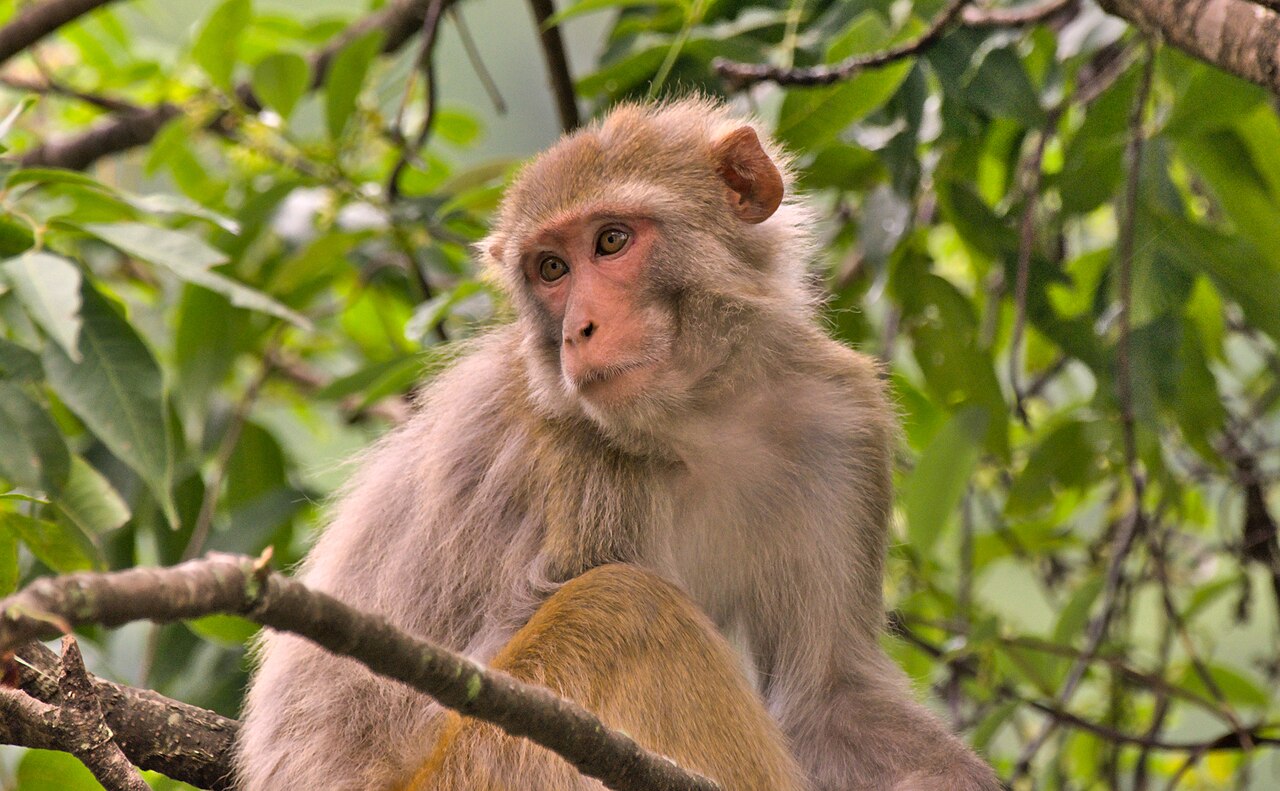
Female Rhesus Macaques That Watched Unfamiliar Female Faces Longer Were More Likely to Have Offspring the Following Year
At a semi-free-ranging population in Puerto Rico, females that spent longer looking at an unfamiliar female’s neutral face…
Read story →
Human Activity and Large Wildlife Overlapped Most in Chitwan’s Buffer Zone
Camera traps across Chitwan National Park and surrounding forests showed the greatest overlap of human activity with large…
Read story →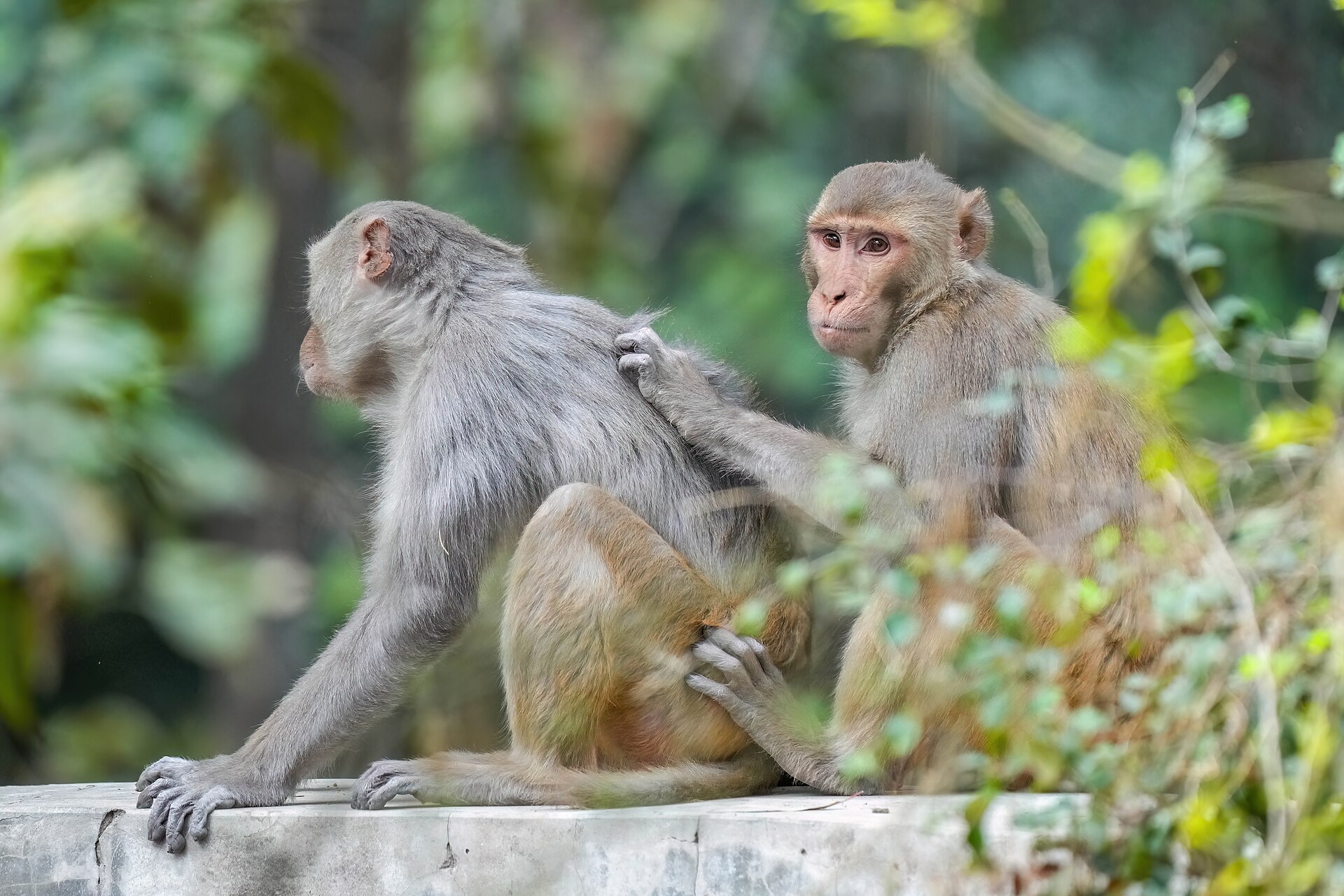
Rhesus Macaques Made More Accurate Choices When They Responded Faster
Rhesus macaques choosing between two alternatives were more accurate when they responded faster, even after choice difficulty was…
Read story →SPECIES IN THIS STORY
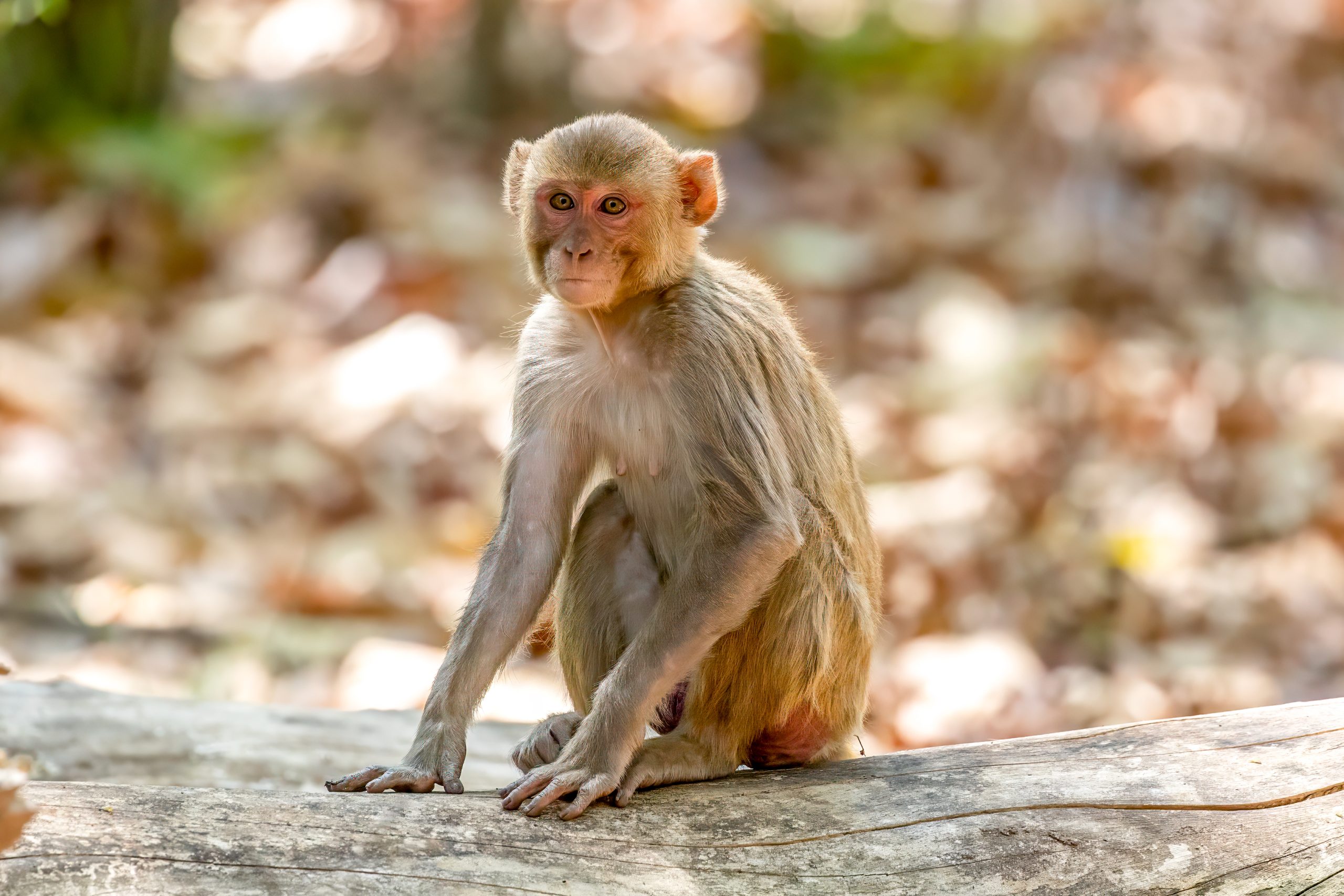
Species in this story
Independent research and conservation news archive
Did you find this information useful?
Wildlife Vagabond is independently built and maintained. Voluntary support helps cover source verification, hosting and continued work on research and conservation news.
Support Wildlife VagabondThe news archive will remain freely available.