
Amur Tiger IL-15 Reduced FPV-Induced Stress and Apoptosis in Cultured Feline Cells
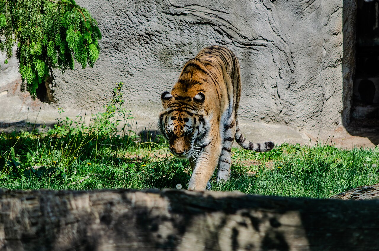
Researchers cloned and compared six cytokines from Amur tigers (Panthera tigris altaica). Cytokines are signalling proteins that coordinate immune-cell activity, and interleukin-15 (IL-15) produced the strongest sustained stimulation of tiger lymphocytes—white blood cells involved in immune responses—among the molecules tested. They then used recombinant Amur tiger IL-15—a laboratory-produced protein made from the cloned tiger gene—in cultured feline kidney cells exposed to feline panleukopenia virus (FPV), a parvovirus that can cause severe disease in felids.
Blood was collected from four healthy adult Amur tigers at Heilongjiang Northeast Tiger Park in China. The researchers cloned IL-2, IL-4, IL-9, IL-13, IL-15 and IL-21 and produced recombinant versions of the proteins. Their amino-acid sequences were more than 90% identical to corresponding cytokines in other felids. IL-15 gave the strongest and most sustained lymphocyte-proliferation response at the highest tested concentration and was selected for the virus experiments.
The virus experiment used cultured cat cells, not infected tigers
For the FPV work, the researchers used F81 feline kidney cells. Cells were pretreated with recombinant Amur tiger IL-15 and then challenged with the virus. No tiger was experimentally infected, and the study did not test IL-15 as a treatment in living tigers.
Compared with infected cells that did not receive IL-15, treated cells maintained a more stable mitochondrial membrane potential. Proteins involved in mitochondrial fusion and fission also shifted toward a more stable balance, with increased MFN1 and MFN2 and reduced Drp1.
Oxidative stress and programmed cell death were reduced
IL-15 pretreatment increased expression of the antioxidant enzymes SOD1 and SOD2 and reduced the accumulation of reactive oxygen species. It also shifted the apoptosis pathway toward lower programmed cell death: pro-apoptotic signals including Bax, p53 and caspases declined, while anti-apoptotic Bcl-2 increased. The cellular protective effects remained detectable for up to 48 hours after infection in this experimental system.
The result is therefore a cell-culture finding about how an Amur tiger immune protein can affect FPV-stressed feline cells. The authors present it as comparative information on felid immune biology and a possible starting point for future conservation-medicine research. Demonstrating safety or therapeutic benefit in Amur tigers would require separate in-vivo studies.
Molecular biology & biochemistry
Related news

Human Activity and Large Wildlife Overlapped Most in Chitwan’s Buffer Zone
Camera traps across Chitwan National Park and surrounding forests showed the greatest overlap of human activity with large…
Read story →
India launches a new evaluation cycle for its 58 tiger reserves
India’s sixth management effectiveness evaluation is planned to cover all 58 tiger reserves. The new cycle adds solid…
Read story →
One Male Tiger Confirmed in Bhutan’s Biological Corridor 7
A 5,144-trap-night survey photographed one adult male tiger at two stations in Bhutan’s Biological Corridor 7, confirming use…
Read story →SPECIES IN THIS STORY
Species in this story
Independent research and conservation news archive
Did you find this information useful?
Wildlife Vagabond is independently built and maintained. Voluntary support helps cover source verification, hosting and continued work on research and conservation news.
Support Wildlife VagabondThe news archive will remain freely available.