
Antioxidant Gene Expression Rose in Roe Deer Testes After the Rut

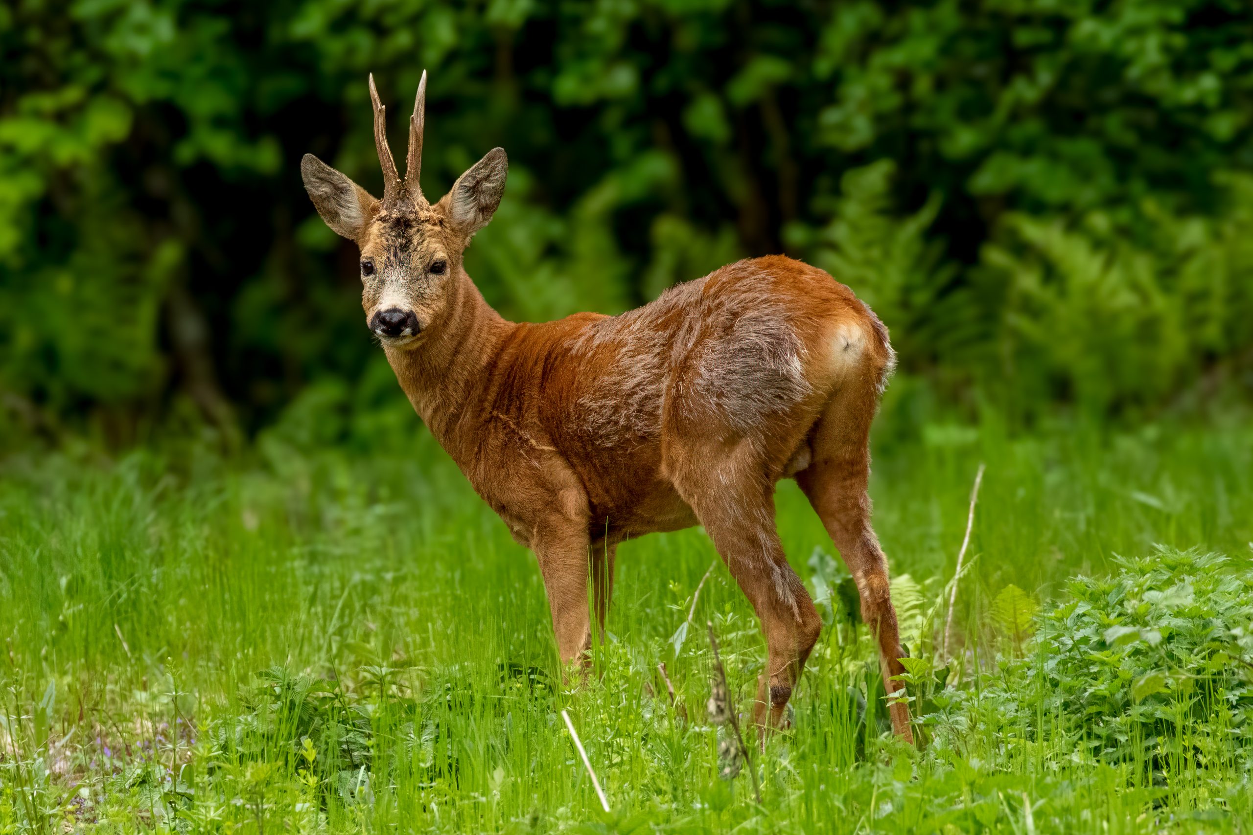
Testicular tissue from male European roe deer (Capreolus capreolus) showed a coordinated molecular shift from the period before the rut to the weeks afterward. A 2026 study found higher expression of several antioxidant genes after the rut, together with changes in genes involved in cholesterol metabolism, energy signalling and angiogenesis — the growth and remodelling of blood vessels that support testicular tissue.
The researchers examined testes from 18 mature bucks legally hunted in the south-western Bologna Apennines in Italy in 2018. Nine were sampled before the rut, from 1 June to 15 July, and nine after it, from 15 August to 30 September. They first screened 84 oxidative-stress genes and 84 angiogenesis-related genes, then quantified selected candidates separately in each animal. For several targets they also measured the corresponding protein, allowing the study to test whether changes in messenger RNA were reflected at protein level.
Antioxidant genes increased while a cholesterol-synthesis gene declined
Three antioxidant-related genes — PRDX4, SCARA3 and SOD3 — had higher mRNA expression after the rut. PRDX4 encodes a peroxiredoxin that helps reduce peroxides such as hydrogen peroxide, while SOD3 encodes an extracellular superoxide dismutase that helps neutralise reactive oxygen species. SCARA3 is a scavenger receptor associated with cellular stress responses. SOD3 showed the largest change in the individual analysis, with mRNA expression rising by about eightfold.
The protein data add an important qualification. SOD3 protein changed in the same direction, but the difference between the pre- and post-rut groups was not statistically supported. The study therefore shows a strong transcriptional increase in SOD3, not an equally well demonstrated increase in SOD3 protein. The authors interpret the broader rise in antioxidant-gene expression as part of testicular recovery following the intense reproductive period.
By contrast, DHCR24 expression declined after the rut. DHCR24 encodes an enzyme that catalyses the final step of cholesterol synthesis and is active in steroid-producing tissues such as the testis. The authors relate the lower expression to the seasonal decline in androgen production and suggest that it may reflect a controlled metabolic slowdown as the testes enter post-rut remodelling. DHCR24 protein could not be evaluated reliably because its concentration fell below the sensitivity of the assay.
Leptin and vascular-regulation genes followed different patterns
LEP, the gene encoding leptin, had lower expression after the rut, and leptin protein was also significantly lower. Leptin links energy reserves with reproductive function in seasonal mammals, so the authors interpret the decrease as part of the post-rut shift in the metabolic–reproductive axis.
THBSII expression also declined. It encodes thrombospondin-2, a protein known to inhibit angiogenesis, although its specific role in spermatogenesis remains poorly understood. NRP2 showed the opposite and more complex pattern: its mRNA increased after the rut, while NRP2 protein declined significantly. NRP2 is a co-receptor involved in several signalling systems, including pathways related to vascular remodelling and reproduction.
The mismatch between NRP2 mRNA and protein is biologically important because it shows that transcription and final protein abundance were not moving together. The authors discuss post-transcriptional regulation — for example reduced translation or altered processing of NRP2 — as possible explanations, but these mechanisms were not tested directly.
Overall, the study does not show one single post-rut switch. Instead, antioxidant defence, steroid-related metabolism, energy signalling and vascular regulation changed in different ways as the testes moved out of the reproductive peak. The authors argue that this molecular reorganisation is consistent with seasonal testicular recovery, while the contrasting mRNA and protein results show why gene expression alone cannot be treated as a direct measure of protein abundance or activity.
Molecular biology & biochemistry
Related news
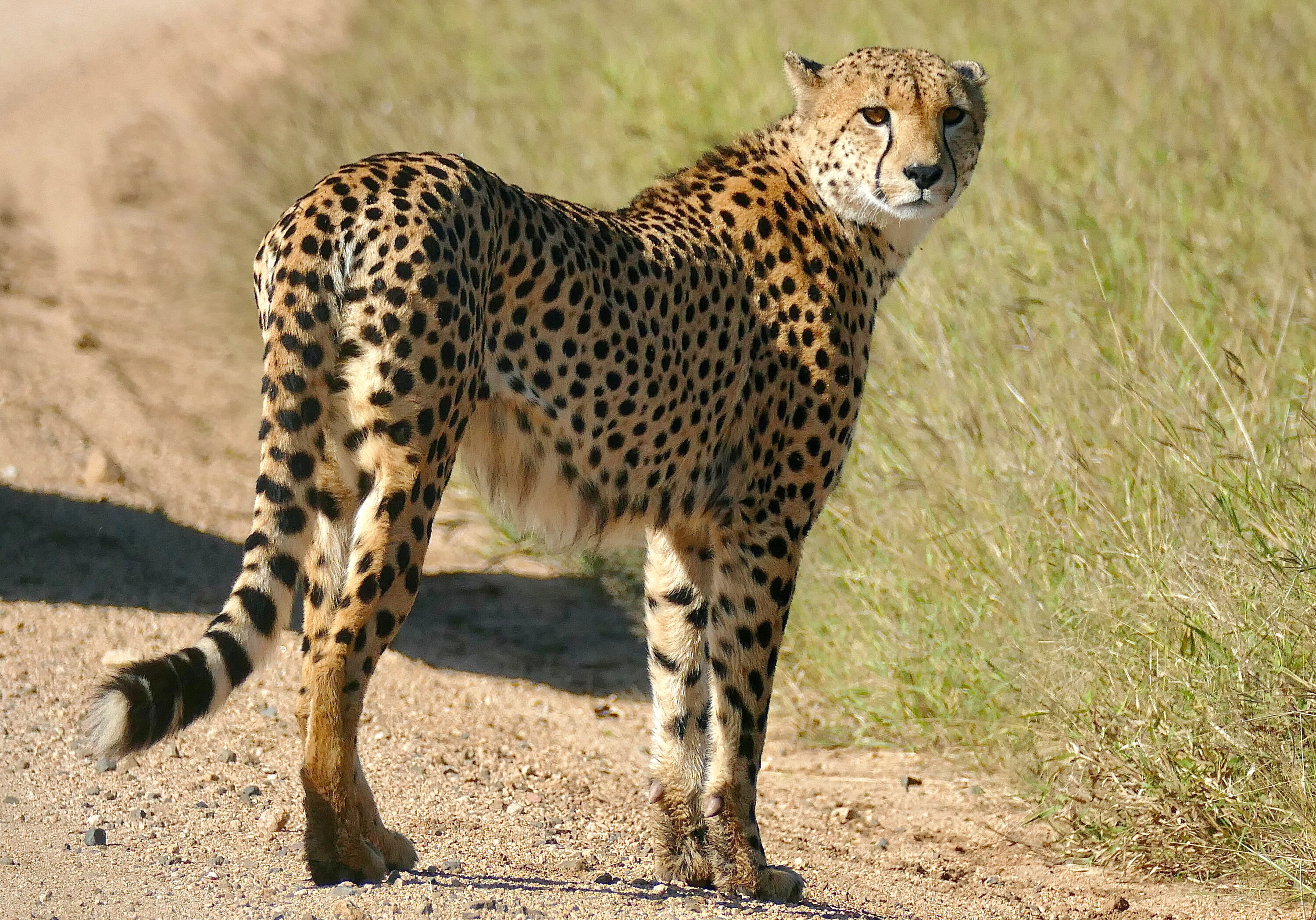
DNA Methylation Shifted as Namibian Male Cheetahs Became Territorial
Paired blood samples from ten free-ranging Namibian males revealed widespread, predominantly increased DNA methylation after the same cheetahs…
Read story →
Svalbard Ptarmigan and Golden Hamsters Shared a TSH Pathway for Seasonal Timing
A comparison of neuroendocrine tissue in summer- and winter-like states found the same core TSH seasonal-timing pathway in…
Read story →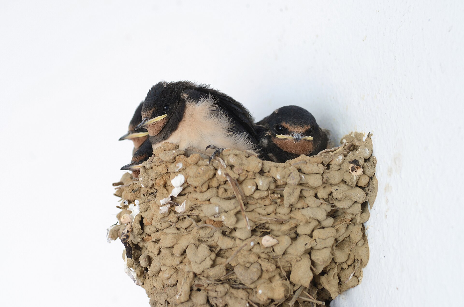
Larger Broods and Older Social Fathers Were Linked to Shorter Telomeres in Male Barn Swallow Nestlings
A long-term study near Milan found shorter telomeres in male barn swallow nestlings from larger broods or with…
Read story →SPECIES IN THIS STORY
Species in this story
Independent research and conservation news archive
Did you find this information useful?
Wildlife Vagabond is independently built and maintained. Voluntary support helps cover source verification, hosting and continued work on research and conservation news.
Support Wildlife VagabondThe news archive will remain freely available.