
Great Spotted Woodpecker Eyes Combined Reinforced Structures with Distinct Injury-related Protein Patterns
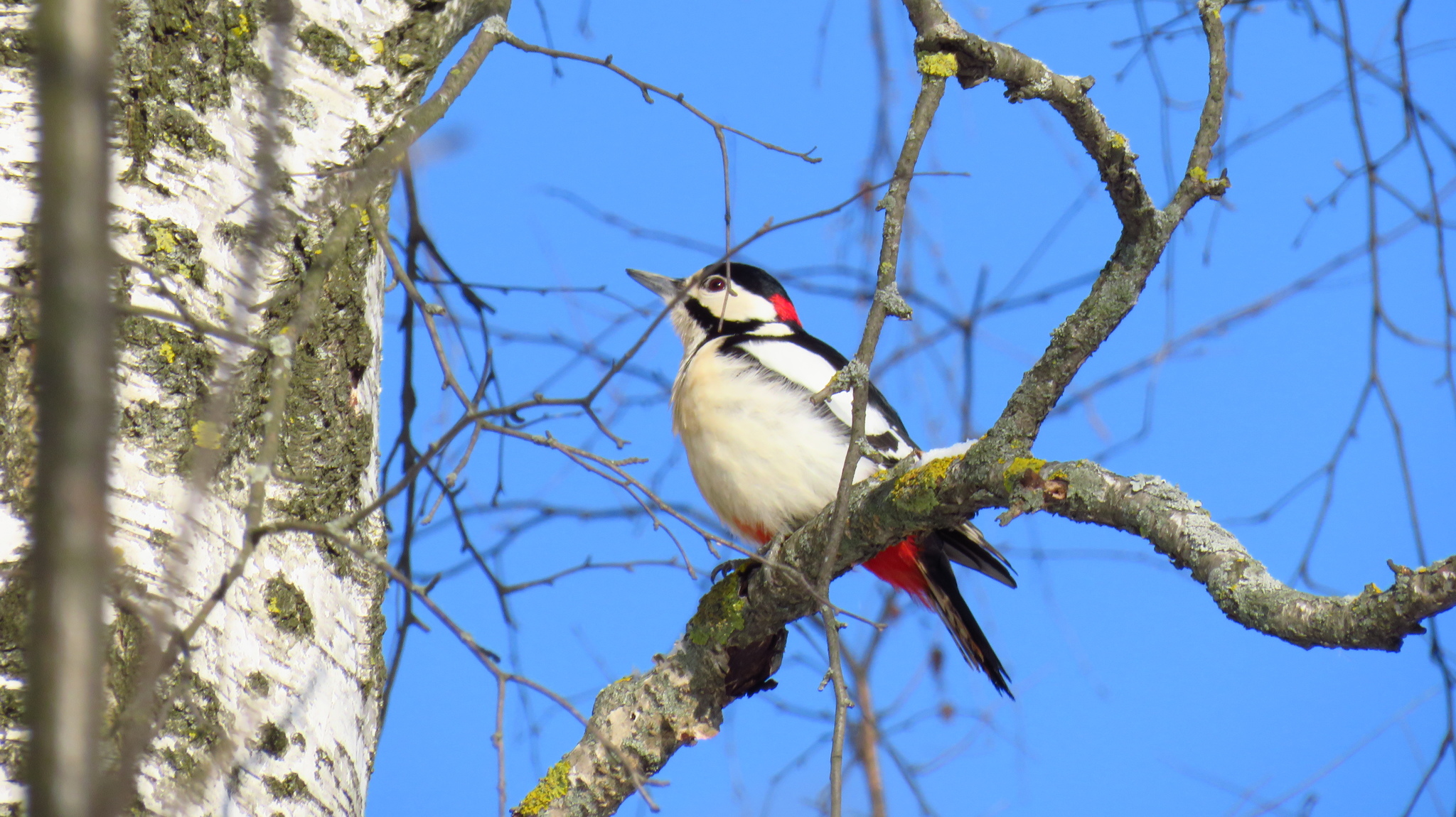
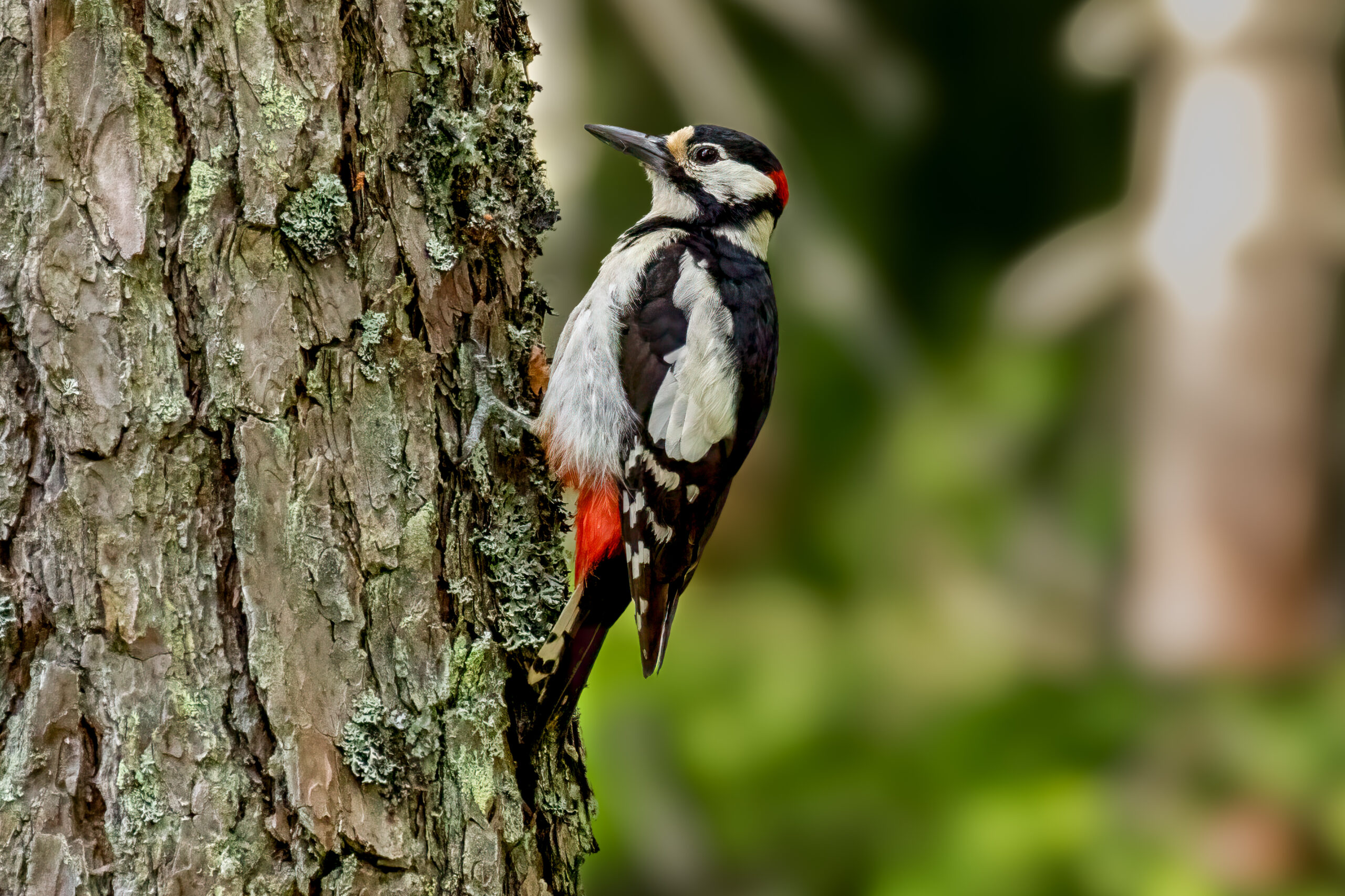
A 2026 study of great spotted woodpecker (Dendrocopos major) eyes found two kinds of features that the authors link to protection during repeated high-acceleration pecking: reinforced eye structures and distinctive patterns of proteins involved in nerve-cell injury and stress responses. Eurasian hoopoe (Upupa epops) served as the comparison species.
The researchers examined frozen eye sections and used Nissl staining to make the organisation of neural tissue visible. They also used immunofluorescence, in which fluorescently labelled antibodies reveal where particular proteins occur in the tissue. This allowed them to compare both eye anatomy and the distribution of β-APP, p-Tau, p-CRMP2, HSP70 and mTOR.
A reinforced outer wall and a more complex pecten
Great spotted woodpeckers had a denser, partly ossified sclera than the hoopoes. The sclera is the tough outer wall of the eye, so additional mineralisation can provide greater structural support during rapid acceleration and deceleration.
The woodpecker eye also had a more complex pecten. The pecten is a folded, highly vascular structure that projects into the vitreous body of a bird’s eye and helps support the retina metabolically. The authors interpret the more elaborate pecten together with the reinforced sclera as the structural layer of the woodpecker’s protection against repeated pecking impacts.
Protein patterns pointed to a second, cellular layer
The molecular results centred on proteins associated with axons, cellular stress and responses to neural injury. β-APP, or beta-amyloid precursor protein, can accumulate when transport along nerve-cell axons is disrupted and is therefore widely used as a marker of axonal injury. In the woodpecker eye it was found mainly in the outer nuclear layer of the retina, the layer containing the nuclei of photoreceptor cells.
Phosphorylated CRMP2 (p-CRMP2) was concentrated mainly in the ganglion cell layer. CRMP2 is involved in axon growth and cytoskeletal organisation, while retinal ganglion cells send their axons into the optic nerve. HSP70, a stress-response protein that helps cells handle damaged or misfolded proteins, occurred in the same locations as phosphorylated tau (p-Tau), a protein form associated with changes to the neuronal cytoskeleton after injury. mTOR, a signalling protein involved in cell growth, metabolism and repair responses, showed different expression patterns that the authors related to different stages of injury response.
The molecular mechanism is an interpretation of tissue patterns
From these distributions, the authors propose that regulation involving CRMP2, HSP70 and mTOR helps prevent abnormal accumulation of β-APP and p-Tau in optic-nerve-related cells and thereby limits damage. The study did not experimentally switch these pathways on or off, so this is a mechanistic interpretation of the observed protein patterns rather than a direct experimental demonstration that any one protein prevents injury.
Together, the reinforced sclera and complex pecten on one hand, and the injury- and stress-related protein patterns on the other, led the authors to describe a two-layered adaptive system in the great spotted woodpecker eye. The work is exploratory, but it provides a biological framework for studying how repeated high acceleration can be tolerated without obvious eye injury.
Physiology
Related news
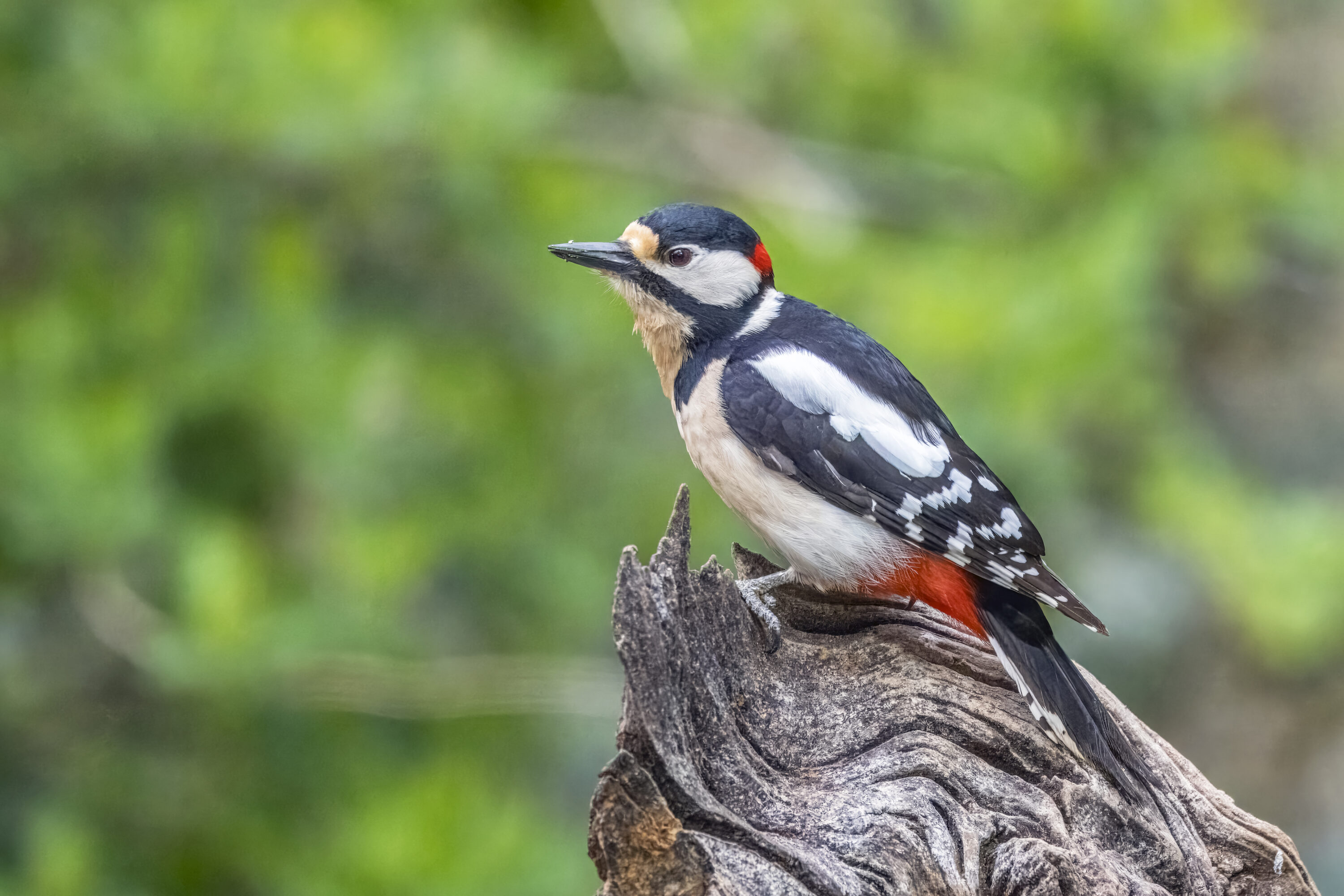
More Woodpeckers in Large, Connected Forests
In the Seoul Metropolitan Area, researchers found more Great Spotted and Japanese Pygmy Woodpeckers in large forest patches…
Read story →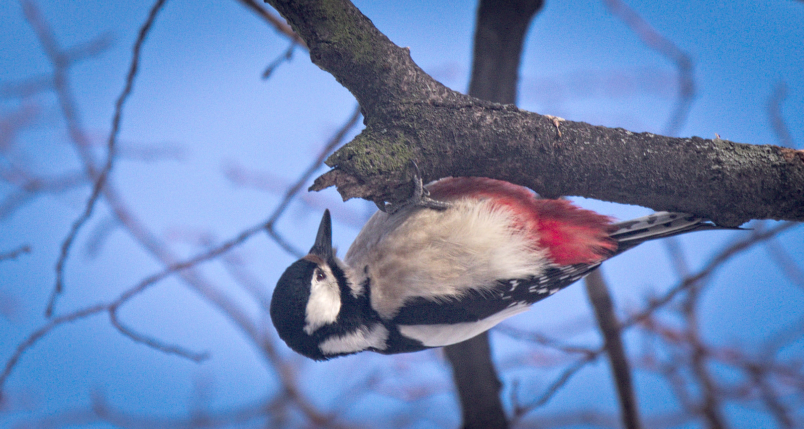
Six European Woodpeckers Shared a Common Eyelid Plan but Showed Microscopic Variation
Microscopic study of 40 woodpeckers from six European species found the same basic eyelid plan, including a fibrous…
Read story →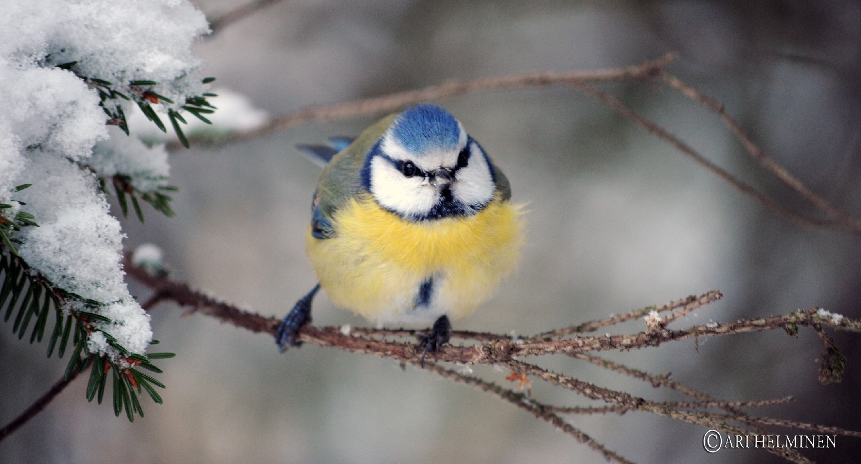
Mean Eurasian Blue Tit Winter Count Rose Fourfold in Finnish Urban Survey Plots
Across 31 Finnish settlements, mean Eurasian blue tit counts rose from 2.8 birds per standardised urban plot in…
Read story →SPECIES IN THIS STORY
Species in this story
Independent research and conservation news archive
Did you find this information useful?
Wildlife Vagabond is independently built and maintained. Voluntary support helps cover source verification, hosting and continued work on research and conservation news.
Support Wildlife VagabondThe news archive will remain freely available.