
Six European Woodpeckers Shared a Common Eyelid Plan but Showed Microscopic Variation
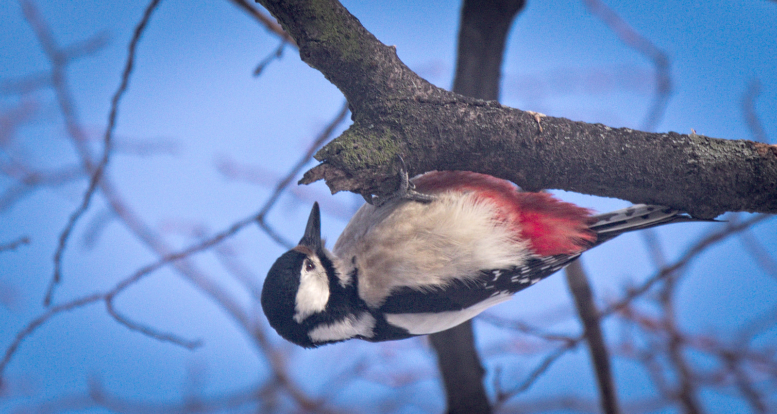
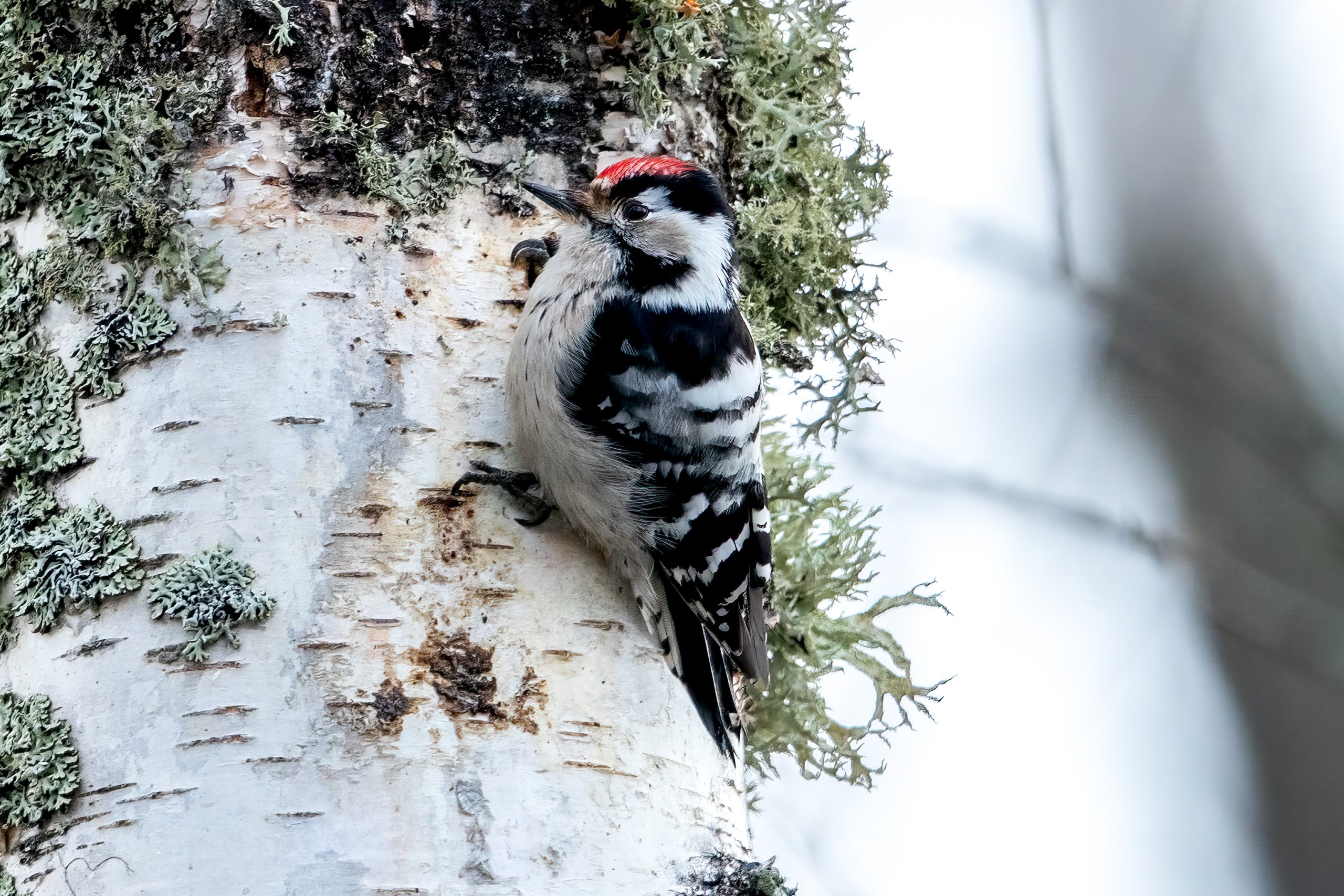
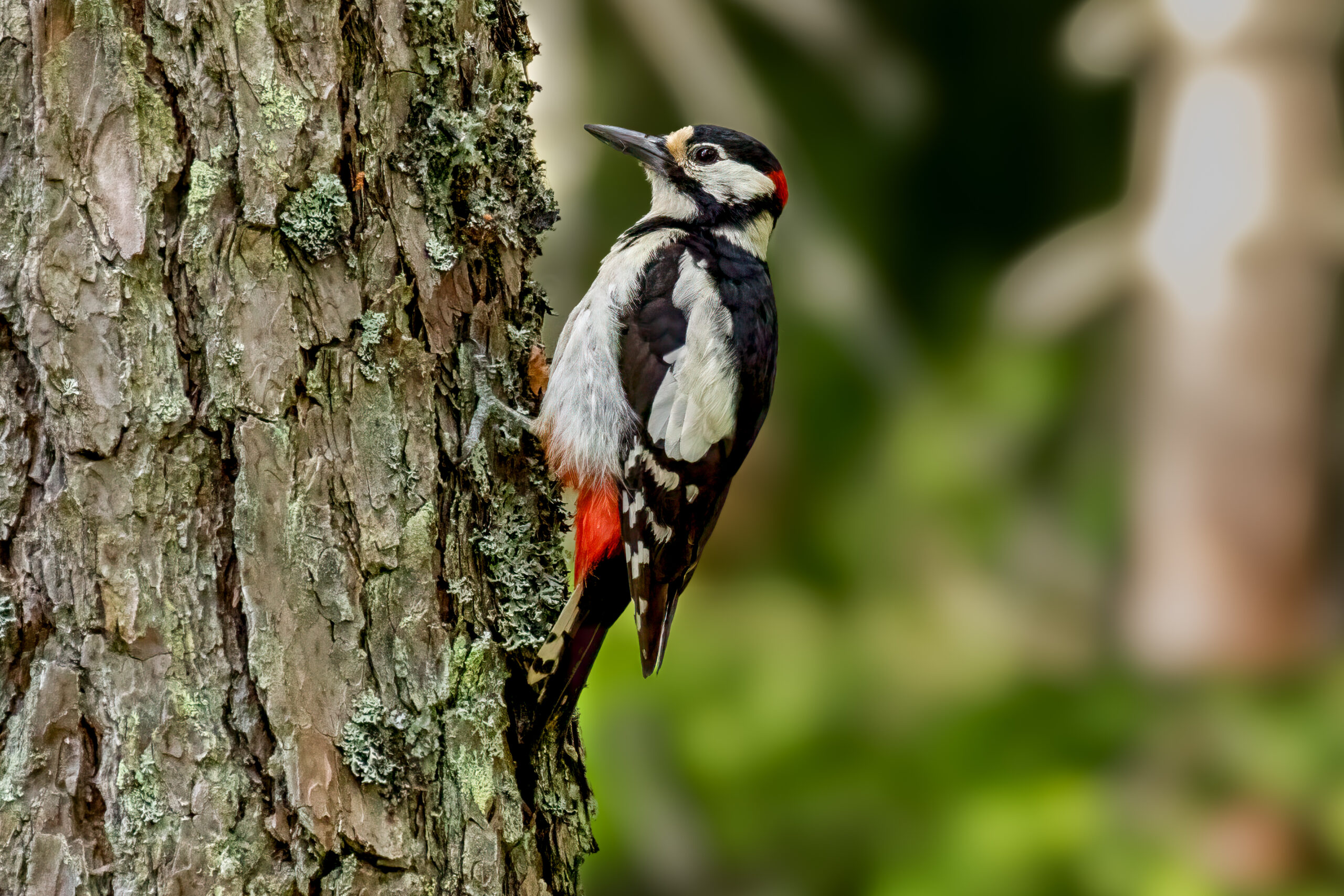
A Polish comparative anatomical study of six European woodpeckers found the same basic microscopic organization in the upper and lower eyelids, alongside descriptive variation in epithelial thickness, skin folds, melanin distribution and parts of the nictitating membrane. The study included gray-headed woodpecker (Picus canus), European green woodpecker (Picus viridis), black woodpecker (Dryocopus martius), great spotted woodpecker (Dendrocopos major), middle spotted woodpecker (Dendrocoptes medius) and lesser spotted woodpecker (Dryobates minor).
The researchers examined the upper eyelid, lower eyelid and nictitating membrane — the mobile third eyelid that can sweep across the eye — in 40 wild woodpeckers found dead in forest and parkland habitats between 2020 and 2023. Great spotted woodpecker accounted for 18 birds and European green woodpecker for 17; lesser spotted woodpecker was represented by two, and gray-headed, black and middle spotted woodpeckers by one each. The tissue was studied with routine histology—microscopic examination of thin tissue sections—and histochemical stains that make particular tissue components easier to distinguish.
The lower eyelid combined mechanical support with local immune tissue
Across all six species, the outer eyelid surface was keratinized and the conjunctival surface contained mucus-producing goblet cells. The lower eyelid contained a compact fibrous tarsal plate that provides structural support. It also contained conjunctiva-associated lymphoid tissue (CALT), made up of lymphoid nodules or aggregates and diffuse lymphocytes. The authors interpret this tissue as a possible site of local immune surveillance on the ocular surface.
The nictitating membrane had a folded organization, with regional variation in epithelial structure, pigmentation and the shape of its leading edge and folds. Together with the eyelids, these structures could contribute to tear-film distribution, removal of small particles and mechanical protection when woodpeckers climb, drill and forage on tree trunks.
Small samples make the species differences tentative
The study provides the first comparative microscopic baseline for these ocular structures in European woodpeckers, but the authors are careful not to turn the observed differences into fixed species traits. The strongly unequal sample sizes — especially the species represented by only one or two birds — mean the interspecific differences should be treated as descriptive observations from the examined material.
Anatomy & morphology
Related news
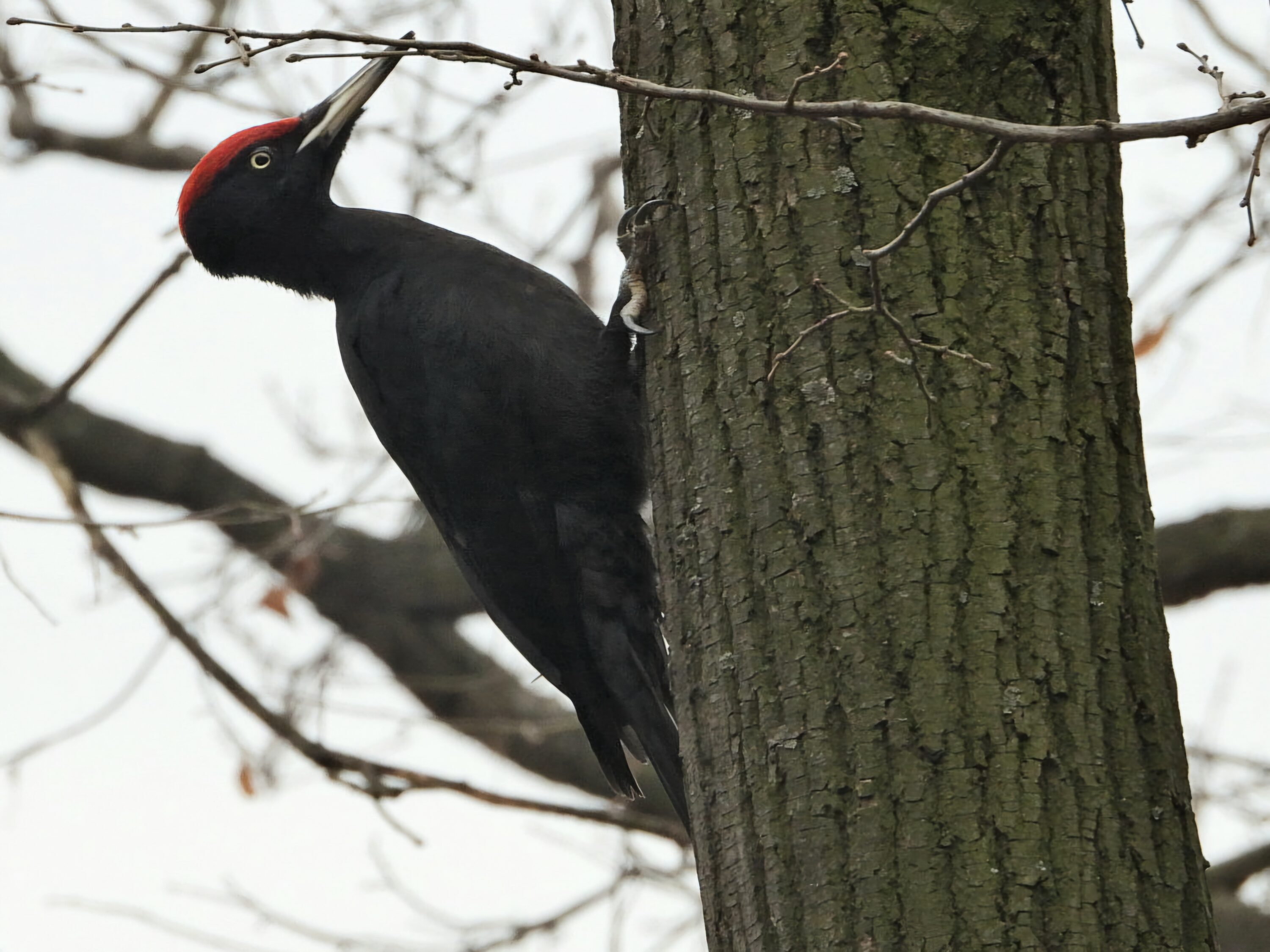
Inside inspections reveal animals missed at Black Woodpecker cavities
In Poland’s Augustów Forest, researchers recorded 50 animal taxa associated with Black Woodpecker cavities. Entrance cameras mostly recorded…
Read story →
More Woodpeckers in Large, Connected Forests
In the Seoul Metropolitan Area, researchers found more Great Spotted and Japanese Pygmy Woodpeckers in large forest patches…
Read story →
Fresh Black Woodpecker Evidence Found at Mount Moriyoshi, but No Detections in Iwate or Shirakami
A 2026 survey found fresh black-woodpecker evidence at Mount Moriyoshi in Akita, while 169 hours of acoustic monitoring…
Read story →SPECIES IN THIS STORY
Species in this story

Independent research and conservation news archive
Did you find this information useful?
Wildlife Vagabond is independently built and maintained. Voluntary support helps cover source verification, hosting and continued work on research and conservation news.
Support Wildlife VagabondThe news archive will remain freely available.