
Young Tissue Stages of a Blood Parasite in Icterine Warblers Resembled a Different Form
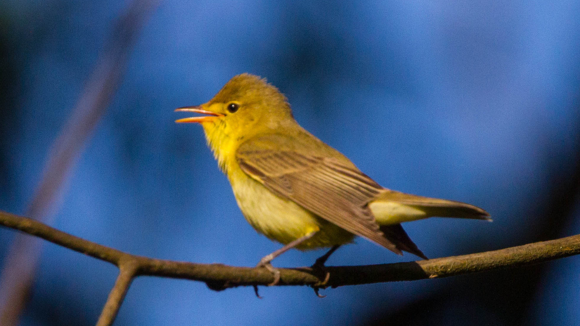
A study of naturally infected icterine warblers (Hippolais icterina) showed that the avian blood parasite Haemoproteus belopolskyi develops only one major form of tissue stage in this host: megalomeronts, large structures in which the parasite multiplies before producing daughter cells called merozoites. Small structures that initially looked like a different stage, known as meronts, were shown by molecular tests to be young megalomeronts of the same parasite.
Haemoproteus are single-celled haemosporidian parasites transmitted between birds by blood-feeding insects such as biting midges. Part of their life cycle occurs in the bird’s tissues before sexual blood stages, or gametocytes, circulate in red blood cells. This multiplication outside the red blood cells is called exo-erythrocytic development or tissue merogony. These tissue stages remain poorly known in most Haemoproteus species and matter biologically because haemoproteid parasites can cause severe disease in bird hosts that are not well adapted to them. This study investigated parasite development rather than clinical disease severity in the warblers.
Twenty-two naturally infected warblers were examined
The researchers studied 22 wild icterine warblers captured at Ventės Ragas Ornithological Station in Lithuania. The birds had first tested positive for Haemoproteus in blood smears. Their organs were then examined with histology and several molecular methods to identify parasite stages within the tissues.
Confirmed H. belopolskyi megalomeronts were found mainly in the kidneys and more occasionally in the heart. Species-specific molecular analyses confirmed megalomeronts in eight birds. Advanced kidney stages were accompanied by local inflammatory reactions, although the authors considered the observed pathology in these naturally infected hosts likely to have had limited overall effect on the birds’ condition.
Young megalomeronts looked like meronts
Meronts and megalomeronts are both tissue stages in which haemosporidian parasites multiply, but mature megalomeronts can become much larger and develop differently from ordinary meronts. In the kidneys, researchers found several small structures that initially had the size and appearance expected of young meronts.
Closer examination showed that these small stages had not begun producing merozoites, the daughter cells characteristic of a mature meront. Instead, their structure formed a developmental series leading into larger, capsule-like megalomeronts and finally to mature megalomeronts filled with merozoites.
The practical implication is that size alone can be misleading: an early megalomeront can resemble a meront before it grows into the much larger form.
Molecular tools confirmed that the stages were the same parasite
Chromogenic in situ hybridisation was used with a species-specific probe. This method attaches a visible molecular signal to parasite genetic material directly inside a tissue section, showing both which parasite is present and exactly where it lies in the organ.
The researchers also used laser capture microdissection to cut individual parasite structures out of tissue sections. DNA from the isolated structures could then be analysed separately. Both the small meront-like stages and the large, advanced megalomeronts were genetically assigned to H. belopolskyi, including the lineage hHIICT1.
This combination of morphology and molecular identification allowed the authors to conclude that the small structures were initial megalomeronts rather than separate meronts.
The life-cycle pattern is simpler than the shapes suggested
Among described Haemoproteus species, some form only meronts, some only megalomeronts and some appear to form both. The new study places H. belopolskyi in the megalomeront-only group in its type vertebrate host, the icterine warbler.
The result also illustrates why tissue-stage morphology alone can be deceptive. Parasites at different ages can vary greatly in size and shape even when they belong to the same developmental pathway. Combining histology with parasite-specific molecular methods therefore provides a more reliable way to reconstruct how these avian blood parasites develop inside their hosts.
Disease, parasites & microbiology
Related news
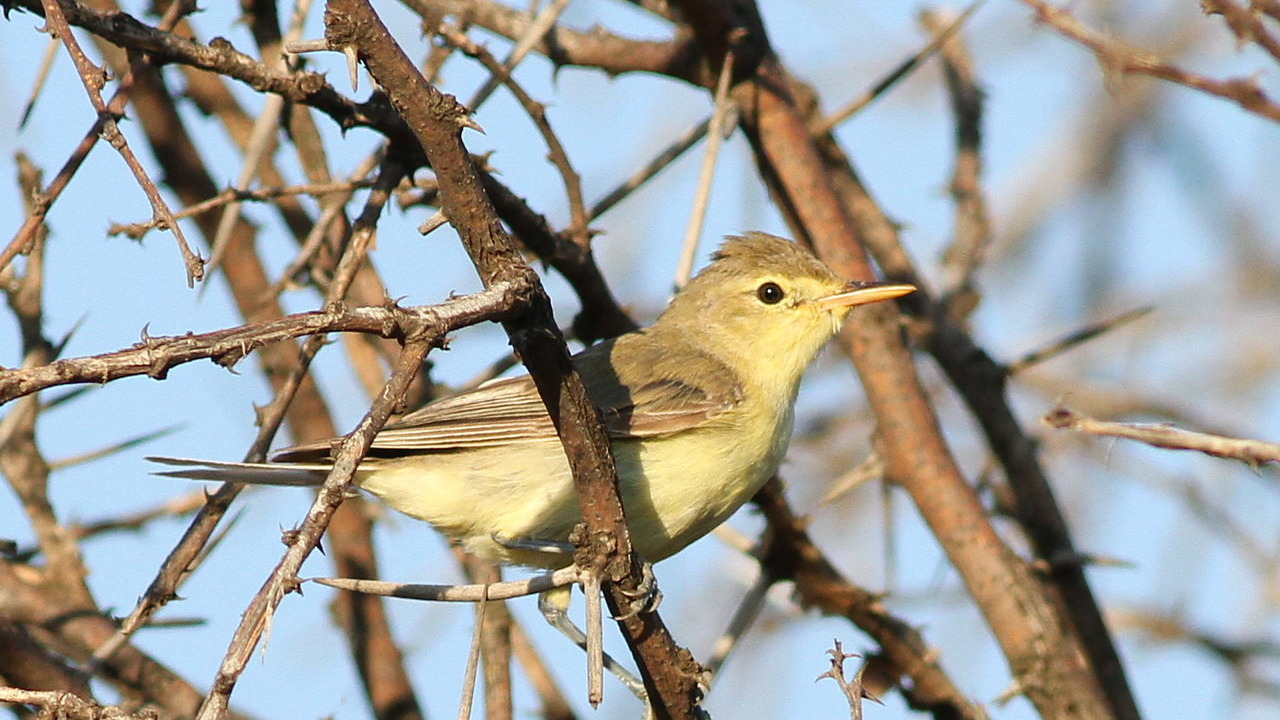
Ten Migrants Followed Early-slow and Late-fast Passage Strategies Through Nigeria
Twenty-four years of ringing data from Jos showed striking species differences in migration timing: Icterine Warblers were among…
Read story →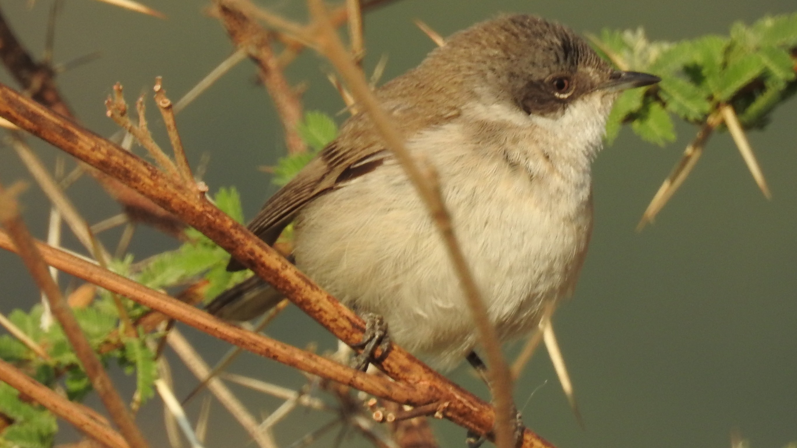
Lesser Whitethroat and Wren Captures Increased While Icterine Warbler Captures Declined at a Hungarian Ringing Site
A standardized 2016–2024 ringing study in southeastern Hungary found significant increases in lesser whitethroat and Eurasian wren captures,…
Read story →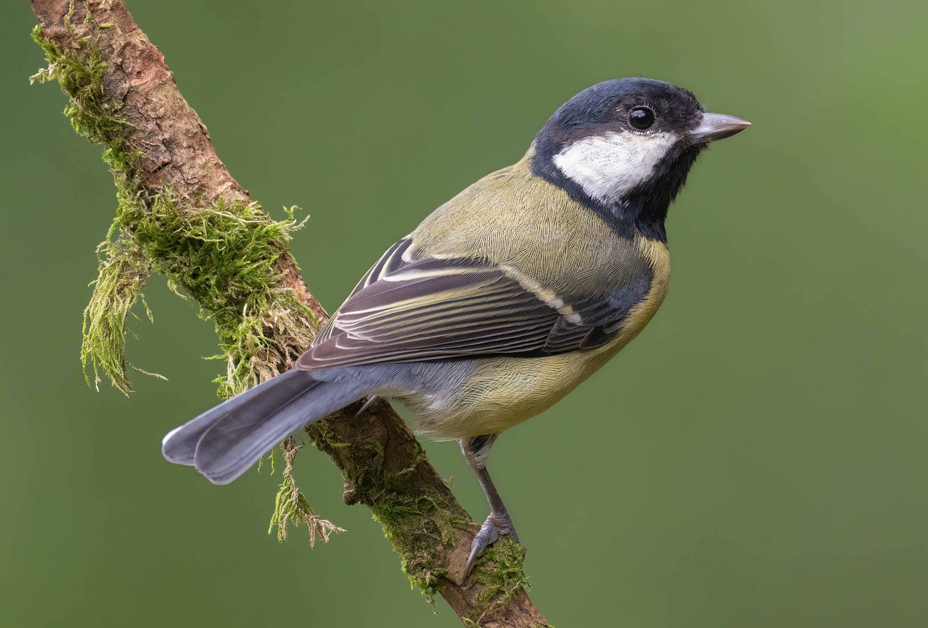
Great Tits Infected Ticks With Borrelia Most Often During Breeding
Across 420 xenodiagnoses in northern Belgium, wild Great Tits were much more likely to transmit Borrelia garinii to…
Read story →SPECIES IN THIS STORY
Species in this story
Independent research and conservation news archive
Did you find this information useful?
Wildlife Vagabond is independently built and maintained. Voluntary support helps cover source verification, hosting and continued work on research and conservation news.
Support Wildlife VagabondThe news archive will remain freely available.