
Human and rhesus macaque immune cells share some signaling patterns—but not all
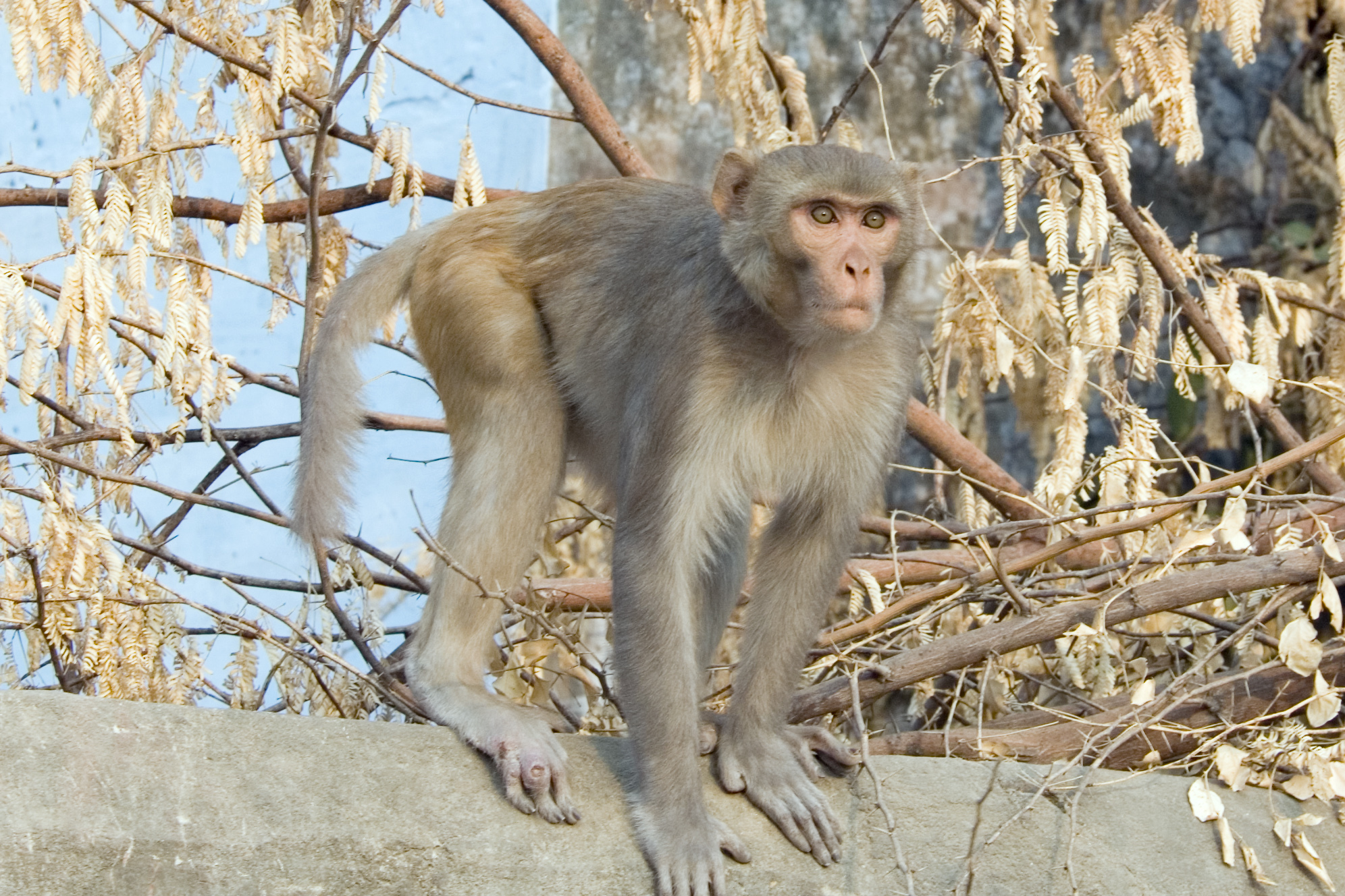
How closely do human and rhesus macaque (Macaca mulatta) immune cells share the same intracellular signaling patterns? A study published as a preprint, not yet peer reviewed, comparing blood cells from the two species combined broad measurements of cell proteins with profiles of signaling activity inside the cells.
Quantum Barcoding (QBC) uses DNA-tagged antibodies to measure proteins on cells. The researchers compared its estimates of immune-cell populations with those from mass cytometry, another method for measuring protein markers in individual cells. They then used a computational tool called MARIO to match profiles measured separately. The combined profiles covered 174 protein features. Their fusion resolved complementary subsets of immune-cell lineages, bringing cell identity and signaling profiles together in a shared framework.
Methods agreed more closely on human cell populations
QBC and mass cytometry agreed more closely on the estimated frequencies of immune-cell populations in human samples than in rhesus macaque samples. The reported Pearson correlation was 0.92 for humans and 0.55 for macaques. In other words, the two methods gave strongly corresponding estimates in the human data, but only a more moderate match in the macaque data.
Agreement in signaling profiles also depended on the cell lineage. Among unstimulated cells, B-cell profiles had a cross-species Spearman correlation of 0.70. This points to a shared pattern in that lineage, but signaling profiles did not match equally well across all immune cells.
GM-CSF responses depended on the cell type
The researchers also exposed blood-cell samples to granulocyte-macrophage colony-stimulating factor (GM-CSF), a cytokine that helps regulate the development and activity of myeloid immune cells. They measured pSTAT5—the phosphorylated form of the STAT5 signaling protein, a transcription factor that can influence gene expression when activated. Increased pSTAT5 thus indicates greater activity in this signaling pathway. After stimulation, pSTAT5 increased in human monocytes and dendritic-cell populations, and in rhesus macaque dendritic cells. The reported increase in macaques was in dendritic cells, not monocytes.
Network analysis inferred similar changes centered on pSTAT5 in human monocytes and rhesus macaque dendritic cells. In both cell groups, the inferred network included a reversal in a pSTAT1 connection and a new coupling between pSTAT3 and pERK. Like STAT5, STAT1 and STAT3 are transcription factors involved in cell signaling, whereas ERK is a protein kinase. The network describes relationships inferred from the measured activity, not necessarily direct molecular binding.
Together, the findings show how broad protein profiles can be linked to signaling measurements across species, while also revealing differences between cell populations. How similar human and macaque immune cells appear therefore depends on which cells and signals are examined.
Molecular biology & biochemistry
Related news
Rhesus macaque vaginal microbiomes changed little across menstrual cycles
Across three cycles in six rhesus macaques, most circulating immune-cell populations and the vaginal microbiome changed little. The…
Read story →
A T-cell vaccine did not improve protection against SHIV, but was linked to better control after infection
In rhesus macaques, adding a T-cell vaccine to PGT121 did not improve protection against SHIV infection. Among animals…
Read story →
Macaques Reacted Slowest Where Either Hand Was Equally Likely to Be Chosen
In a reaching task, two rhesus macaques were slowest to respond near the point where either hand was…
Read story →SPECIES IN THIS STORY
Species in this story
Independent research and conservation news archive
Did you find this information useful?
Wildlife Vagabond is independently built and maintained. Voluntary support helps cover source verification, hosting and continued work on research and conservation news.
Support Wildlife VagabondThe news archive will remain freely available.