
Study Maps the Eye Glands of Natal Red Duikers and Kirk’s Dik-Diks
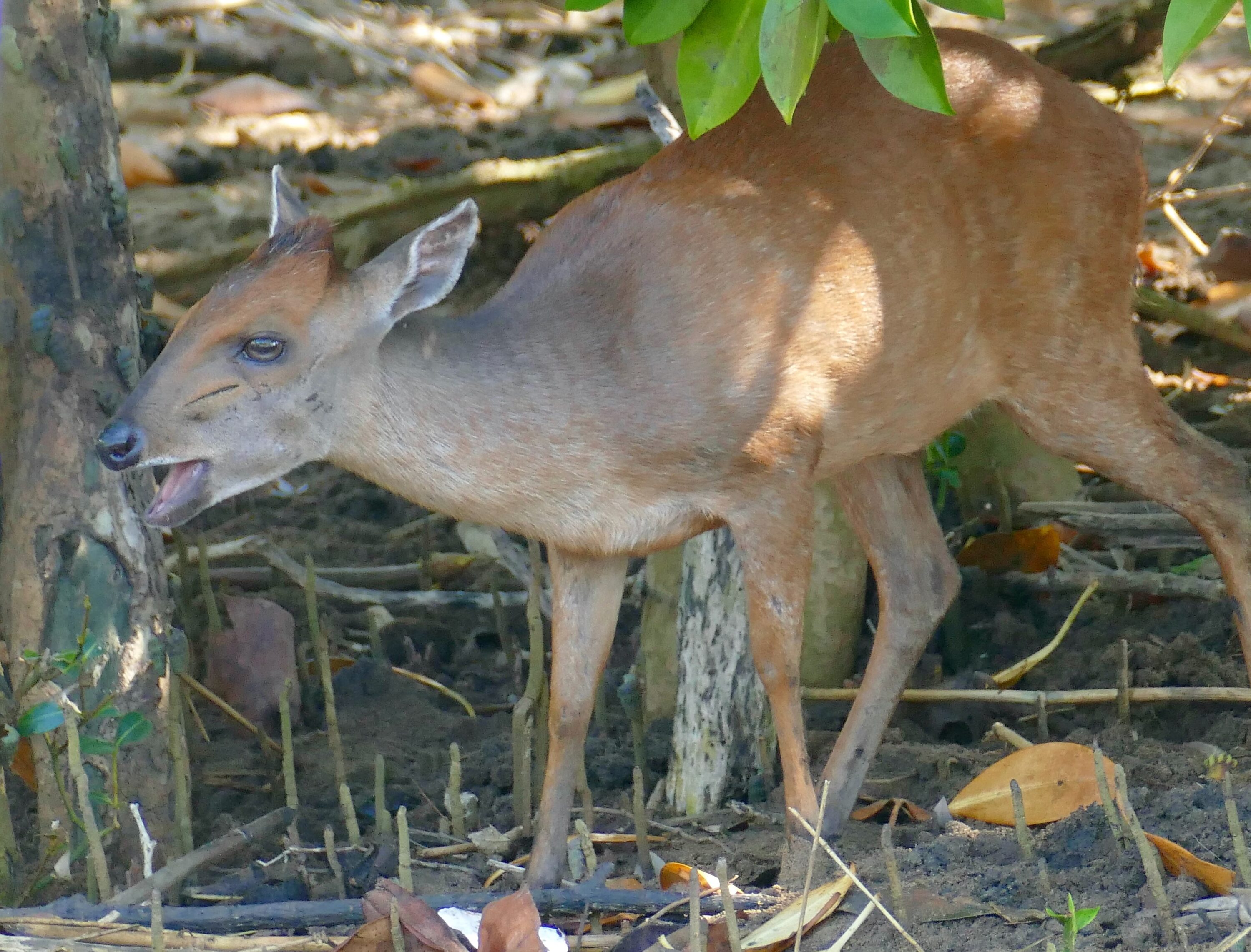
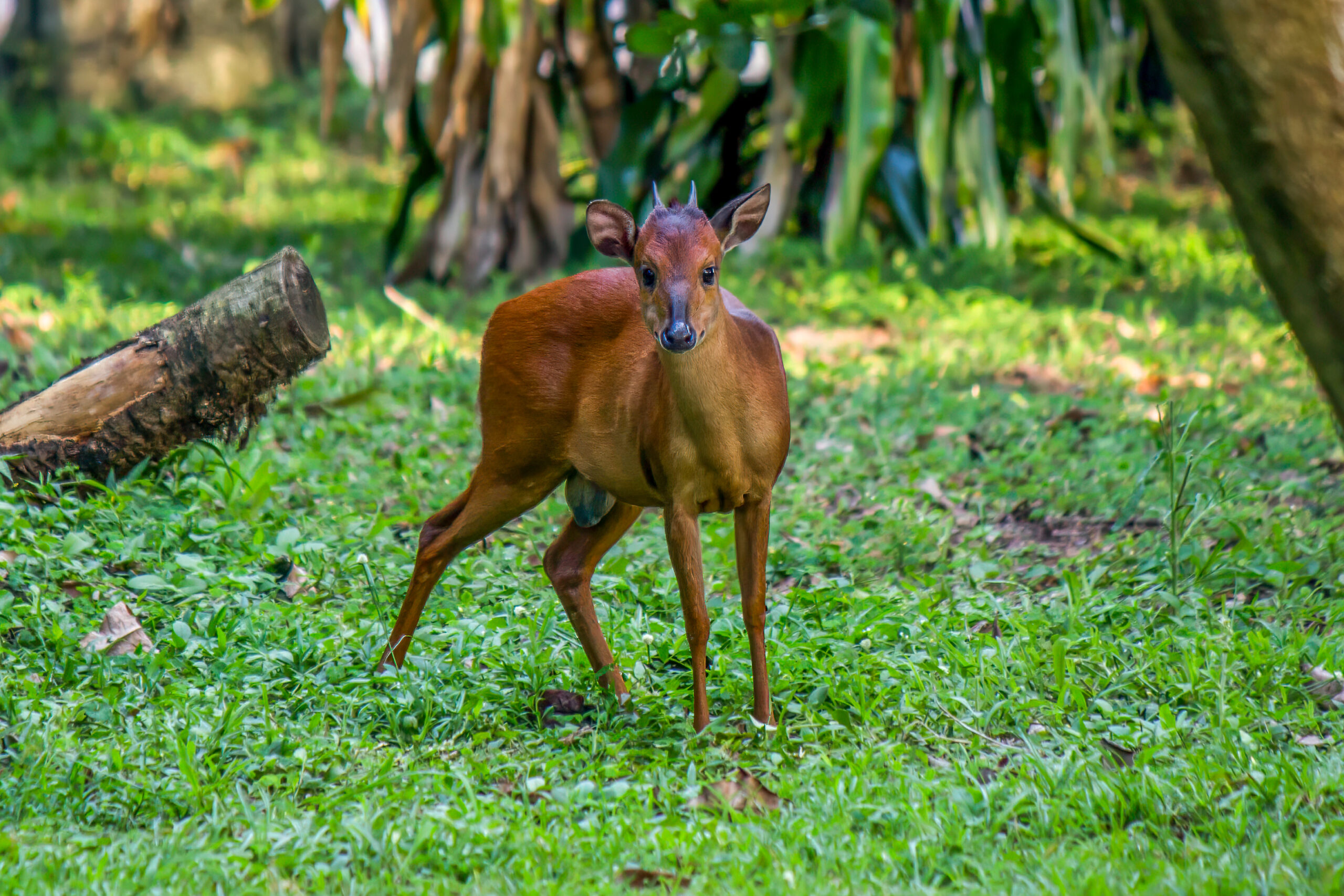
A 2026 anatomical study provides a detailed description of the gland complex that protects the eye surface in Natal red duikers (Cephalophorus natalensis) and compares it with Kirk’s dik-diks (Madoqua kirkii). These structures had been poorly documented in both small African antelopes.
The study examined post-mortem material from five young Natal red duikers and four young Kirk’s dik-diks from Wrocław Zoo in Poland. The animals had died independently of the research, and the researchers reported no visible abnormalities in the eyes or surrounding tissues when the samples were collected.
On both sides of the head, the team examined three glands involved in maintaining and protecting the ocular surface: the Harderian gland, the lacrimal gland and the superficial gland of the third eyelid. The tissues were studied macroscopically and under the microscope, and a series of histochemical stains was used to identify different components of the glandular secretions.
The three glands occupied distinct parts of the orbit
In both species, the three glands occupied distinct parts of the orbit. The Harderian gland was positioned on the inner and lower side of the eye, the lacrimal gland on the upper outer side, and the superficial gland of the third eyelid close to the third eyelid. Microscopically, the glands were built from branching secretory units with tubular and rounded acinar portions, although some were dominated by the rounded acini. The examined glands also differed in capsule thickness, internal septa, lobular organisation and the appearance of the secretory cells.
The histochemical analyses showed a mixture of neutral glycoconjugates and acidic mucosubstances in the secretions, although their relative occurrence varied between glands and between the two groups of animals. Plasma cells and lymphocytes were also present within the glandular tissue, reflecting the immune functions associated with the ocular surface.
The small sample limits species-level conclusions
The authors emphasise that this was a preliminary descriptive study with only five red duikers and four dik-diks. They did not statistically test whether the observed differences between the two small groups were larger than might be expected from individual variation. The observed differences should therefore not be treated as definitive species-level characteristics. Instead, the results establish reference material for comparative anatomy and for veterinary examination of orbital tissues in zoo and wildlife medicine.
Anatomy & morphology
Related news
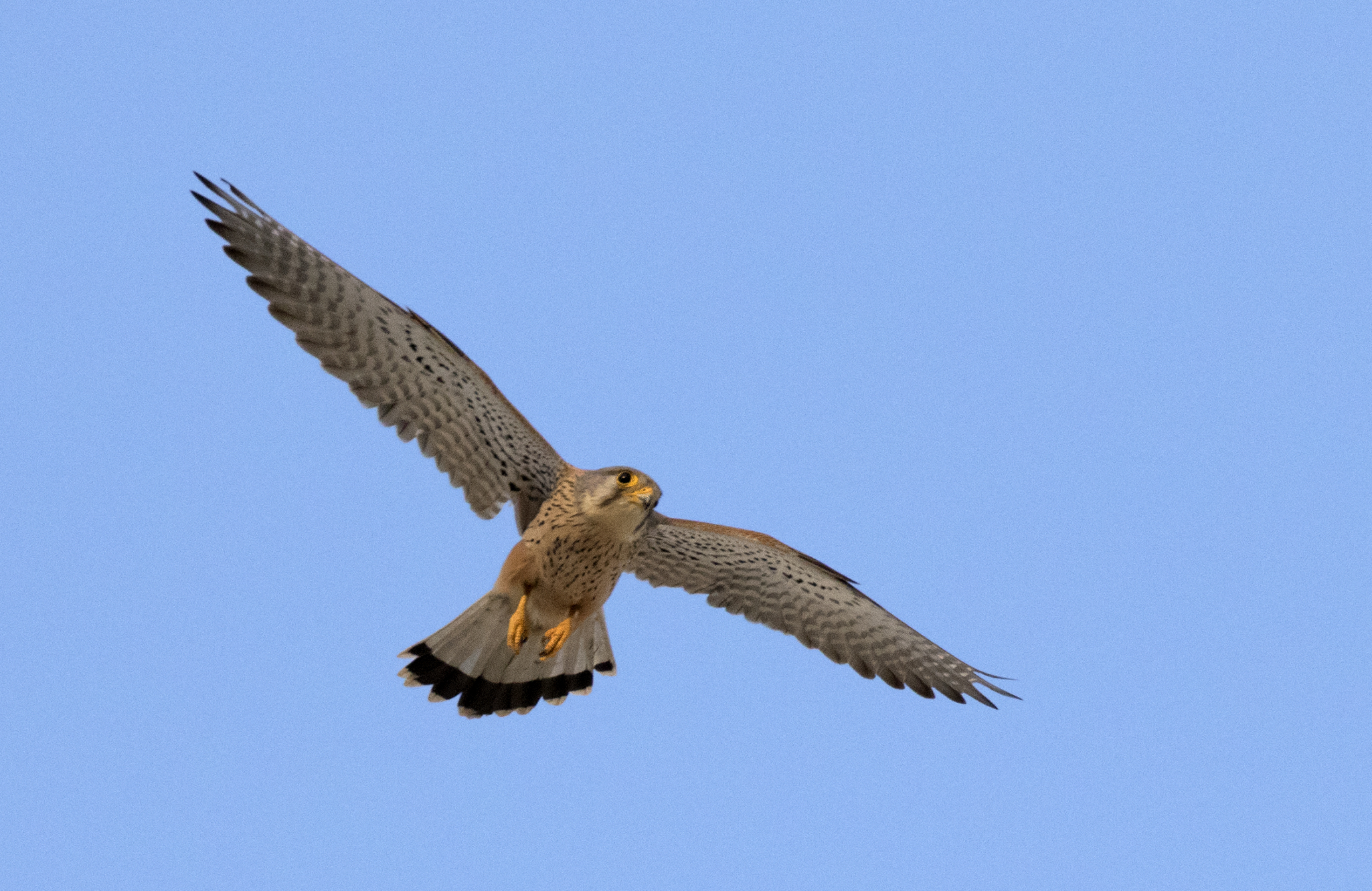
Common Kestrels Had About Twice the Tracheal Mucous-Gland Density of White Storks
A comparative study of three adult males from each of three bird species found 11.32 tracheal mucous glands…
Read story →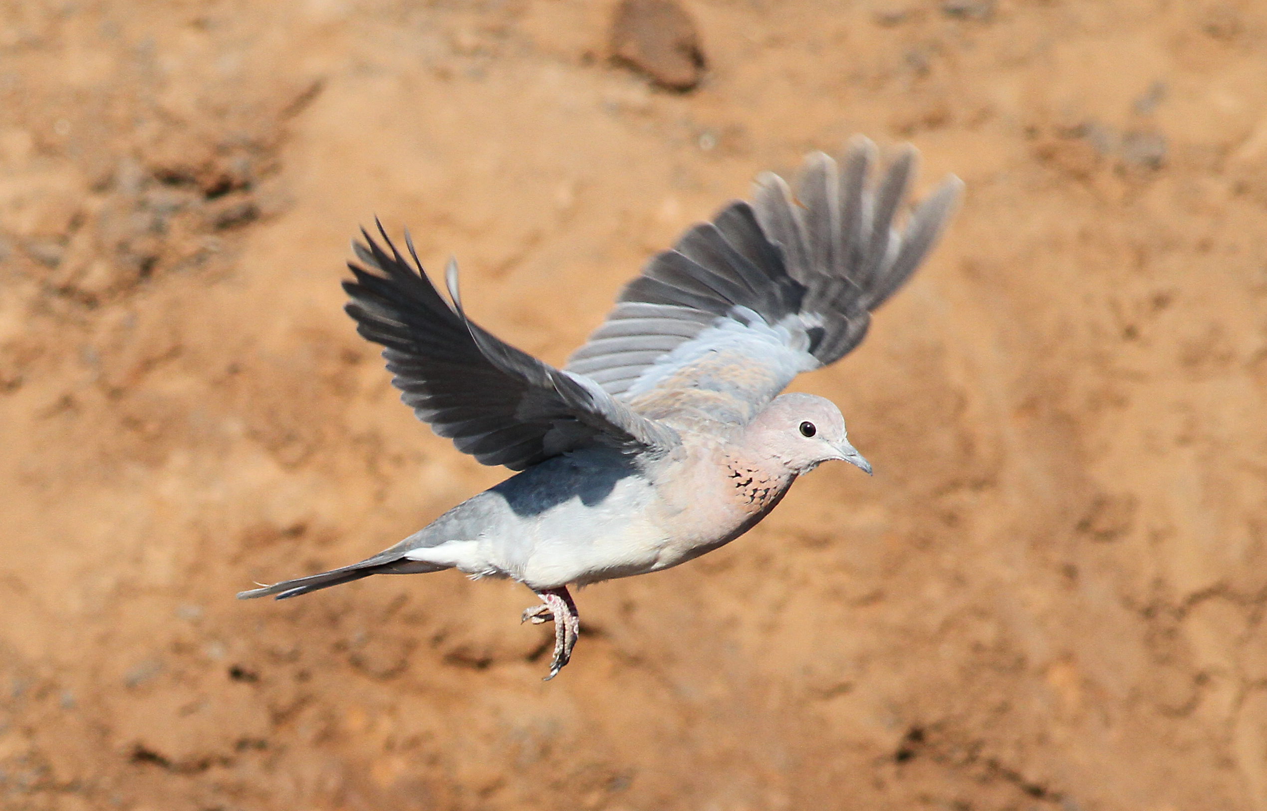
Laughing Dove Eye Pecten Mapped From 12–13 Pleats to Cell Markers
Researchers mapped the pecten oculi, a vascular structure unique to bird eyes, in the laughing dove. Its 12–13…
Read story →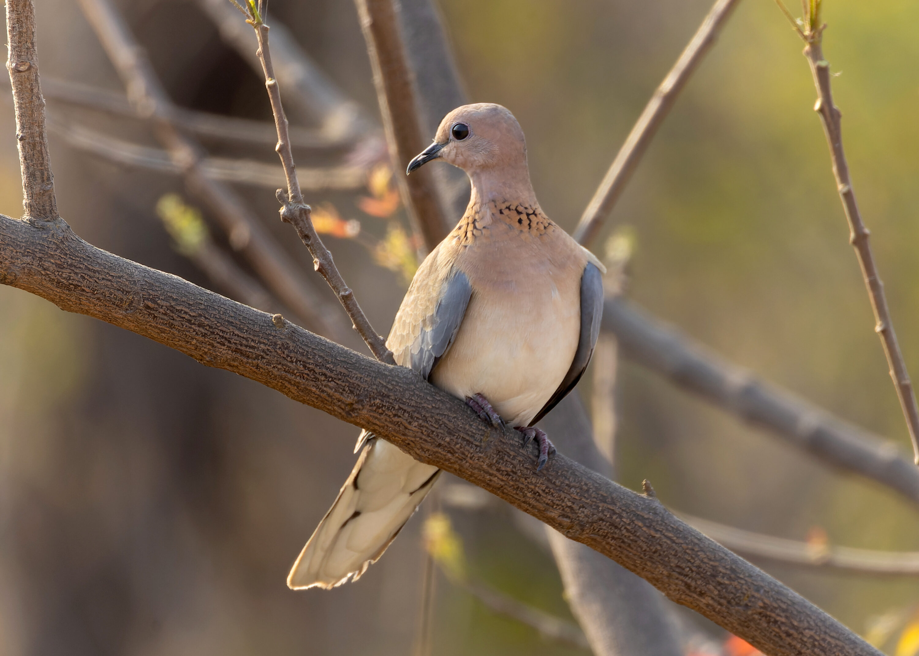
Progressive Greying Recorded in a Laughing Dove in Asia for the First Time
An adult laughing dove photographed in Karnataka, India, had localised white feathers on the head and shoulder region.…
Read story →SPECIES IN THIS STORY
Species in this story
Independent research and conservation news archive
Did you find this information useful?
Wildlife Vagabond is independently built and maintained. Voluntary support helps cover source verification, hosting and continued work on research and conservation news.
Support Wildlife VagabondThe news archive will remain freely available.