
Foot-and-mouth disease virus antigen detected in blood vessels of carrier African buffalo
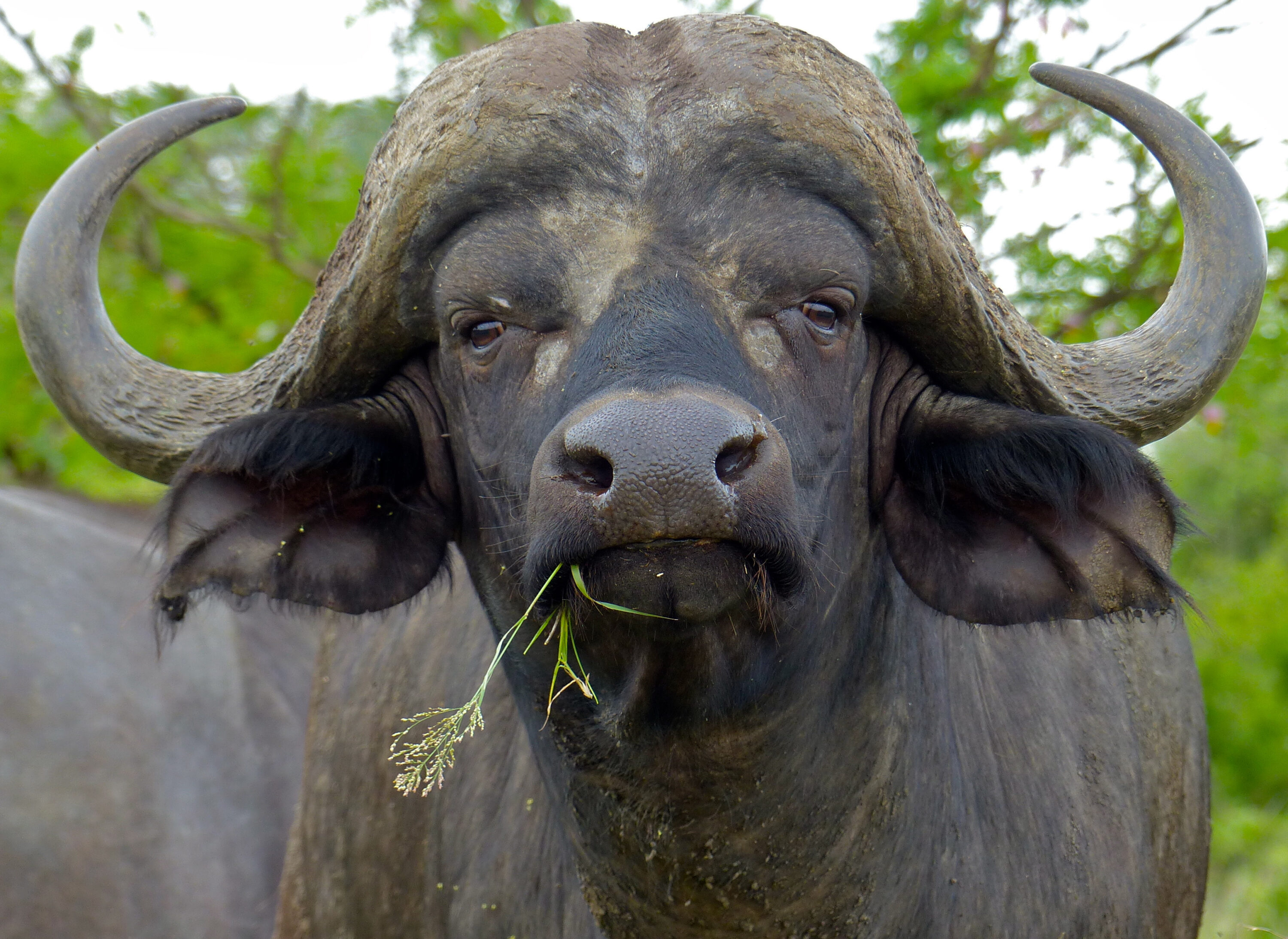
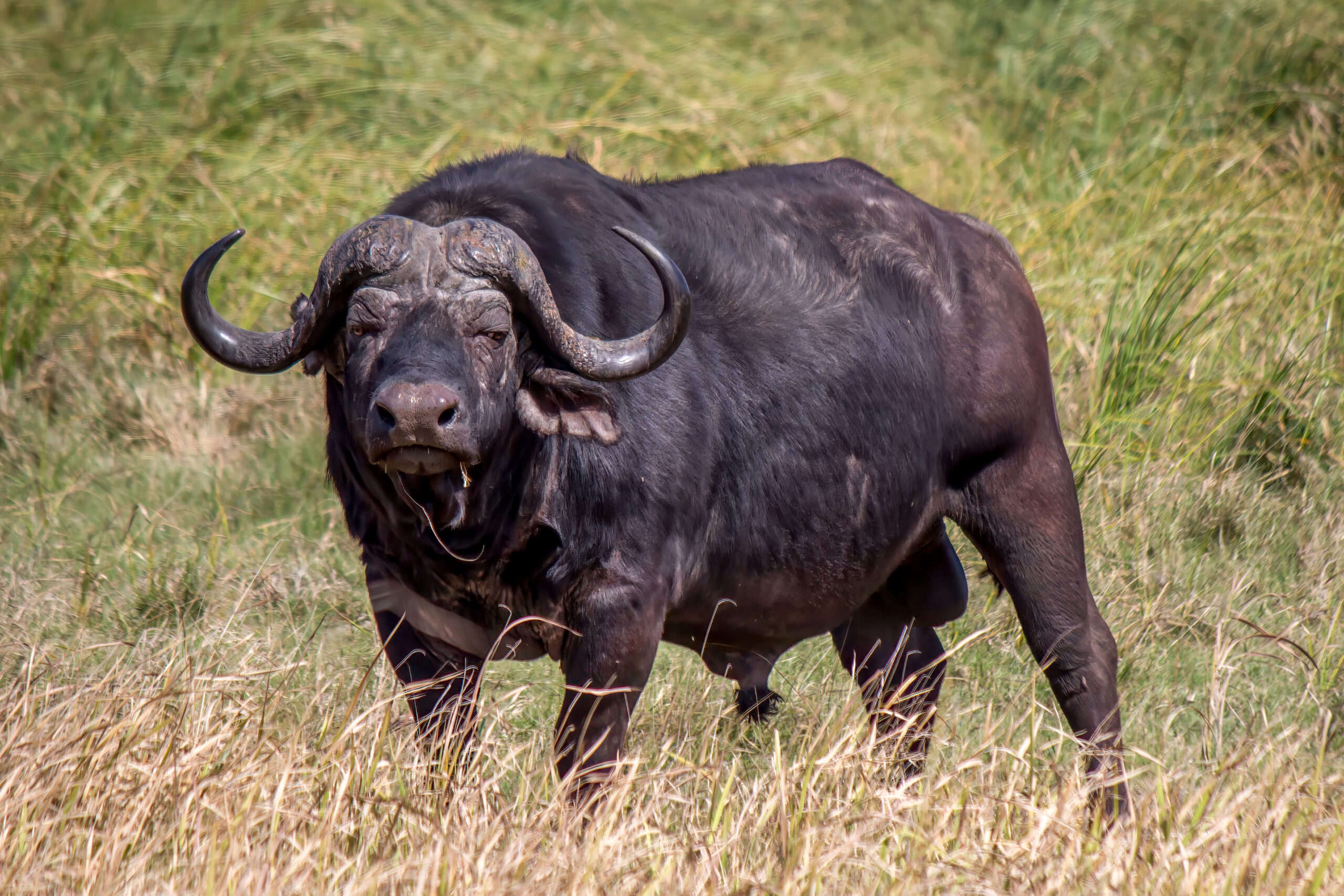
A study published on 13 March 2026 has mapped foot-and-mouth disease virus antigen in tissues from carrier african buffalo (Syncerus caffer). The most prominent staining was found in the skin between the hooves and along the coronary band where the hoof meets the skin, and was mainly associated with the endothelium of small blood vessels. Viral antigen was also detected in the lungs and palatine tonsils.
A carrier animal has recovered from an infection but continues to harbour virus in oropharyngeal fluid — fluid from the mouth and throat region — for more than 28 days after infection. Such an animal is not necessarily contagious. African buffalo can carry the Southern African Territories serotypes of foot-and-mouth disease virus for long periods, but the small amount of virus in carriers makes reliable detection difficult.
The researchers examined formalin-fixed tissues from 15 subadult female buffalo culled for population control in Kruger National Park, South Africa. They compared these with tissues from 15 disease-free buffalo that had repeatedly tested negative while in quarantine. The study animals were between two and four years old.
A PCR test performed around the time of slaughter classified 12 of the 15 Kruger buffalo as positive and three as negative. The team then used immunohistochemistry, or IHC, to make viral proteins visible in tissue sections. In this context, antigen means viral proteins recognised by the antibodies: antibodies against structural proteins of the virus produced coloured staining where antigen was present.
IHC detected antigen in all 15 Kruger buffalo
IHC detected antigen in all 15 Kruger buffalo, including the three that had tested negative by PCR. None of the 15 disease-free control animals showed comparable staining. In the test group, antigen was most often found in the skin between the hooves and along the coronary band where the hoof meets the skin. Positive staining in these areas and in the lungs was mainly associated with the endothelium, the layer of cells lining small blood vessels. The palatine tonsils also contained stained cells, especially within germinal centres involved in immune responses.
No staining was detected in several other tissues, including the tongue, lips, eyelids, ear tips, pharyngeal tissue and retropharyngeal lymph nodes. The distribution differed from previous work that located viral RNA mainly in tonsils and lungs. The authors explain that IHC detects viral proteins, whereas other methods may detect remaining genetic material, so the techniques may highlight different parts or stages of infection.
The three PCR-negative buffalo were still positive by tissue staining
The three IHC-positive but PCR-negative results suggest that tissue staining could help detect low viral loads. The authors describe this as evidence that IHC may offer improved sensitivity under such conditions, but stress that further work is needed to confirm it. The staining was scored manually and semi-quantitatively, and the authors note that a larger sample would be needed to calculate and compare the sensitivity and specificity of IHC with other tissue-based methods.
The results identify vascular tissue as a possible additional site of viral persistence in carrier buffalo. They do not show that these animals were transmitting the virus. The researchers call for larger studies to repeat the result, validate the assay and clarify how carrier animals contribute to foot-and-mouth disease persistence and spread.
Disease, parasites & microbiology
Related news
Food Leaves Its Mark on the Gut Bacteria of Kruger’s Wildlife
Gut bacteria in nine mammal species from Kruger National Park followed clear patterns linked to food and digestion.…
Read story →
Buffalo Gather Around Water and Forest as the Dry Season Arrives
Buffalo herds grew larger and used more forest during the dry season in Ethiopia’s Gambella National Park. Wetlands…
Read story →
First Reported Giardia Detections in Four Wild Herbivore Species in Kenya
Molecular screening of wildlife and livestock in Laikipia produced the first reported Giardia detections in African buffalo, plains…
Read story →SPECIES IN THIS STORY
Species in this story
Independent research and conservation news archive
Did you find this information useful?
Wildlife Vagabond is independently built and maintained. Voluntary support helps cover source verification, hosting and continued work on research and conservation news.
Support Wildlife VagabondThe news archive will remain freely available.